4EYH

Human DNA polymerase iota incorporating dCTP opposite N-(deoxyguanosin-8-yl)-1-aminopyrene lesion


Experimental Data Snapshot

  • Method: X-RAY DIFFRACTION
  • Resolution: 2.90 Å
  • R-Value Free: 0.281 
  • R-Value Work: 0.239 
  • R-Value Observed: 0.242 

wwPDB Validation   3D Report Full Report


Ligand Structure Quality Assessment 


This is version 1.2 of the entry. See complete history


Literature

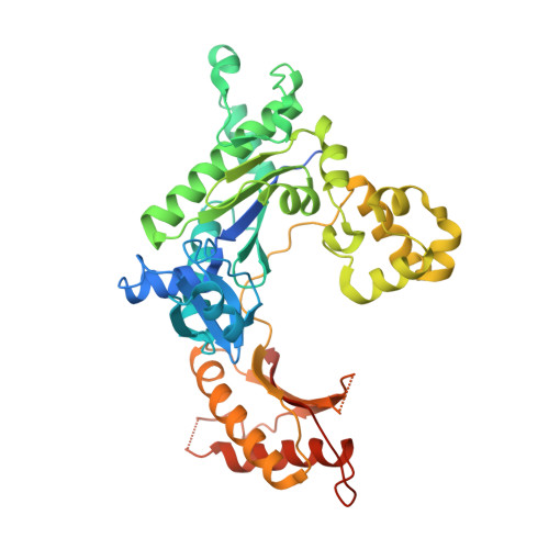
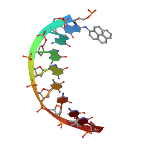
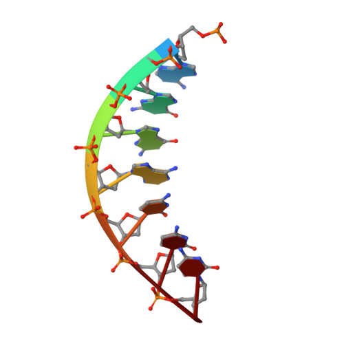
Replication of a carcinogenic nitropyrene DNA lesion by human Y-family DNA polymerase.

Kirouac, K.N.Basu, A.K.Ling, H.

(2013) Nucleic Acids Res 41: 2060-2071

  • DOI: https://doi.org/10.1093/nar/gks1296
  • Primary Citation of Related Structures:  
    4EYH, 4EYI

  • PubMed Abstract: 

    Nitrated polycyclic aromatic hydrocarbons are common environmental pollutants, of which many are mutagenic and carcinogenic. 1-Nitropyrene is the most abundant nitrated polycyclic aromatic hydrocarbon, which causes DNA damage and is carcinogenic in experimental animals. Error-prone translesion synthesis of 1-nitropyrene-derived DNA lesions generates mutations that likely play a role in the etiology of cancer. Here, we report two crystal structures of the human Y-family DNA polymerase iota complexed with the major 1-nitropyrene DNA lesion at the insertion stage, incorporating either dCTP or dATP nucleotide opposite the lesion. Polι maintains the adduct in its active site in two distinct conformations. dCTP forms a Watson-Crick base pair with the adducted guanine and excludes the pyrene ring from the helical DNA, which inhibits replication beyond the lesion. By contrast, the mismatched dATP stacks above the pyrene ring that is intercalated in the helix and achieves a productive conformation for misincorporation. The intra-helical bulky pyrene mimics a base pair in the active site and facilitates adenine misincorporation. By structure-based mutagenesis, we show that the restrictive active site of human polη prevents the intra-helical conformation and A-base misinsertions. This work provides one of the molecular mechanisms for G to T transversions, a signature mutation in human lung cancer.


  • Organizational Affiliation

    Department of Biochemistry, Medical Sciences Building 334, University of Western Ontario, London, ON N6A 5C1, Canada.


Macromolecules

Find similar proteins by:  (by identity cutoff)  |  3D Structure
Entity ID: 1
MoleculeChains Sequence LengthOrganismDetailsImage
DNA polymerase iotaA [auth B]420Homo sapiensMutation(s): 0 
Gene Names: POLIRAD30B
EC: 2.7.7.7
UniProt & NIH Common Fund Data Resources
Find proteins for Q9UNA4 (Homo sapiens)
Explore Q9UNA4 
Go to UniProtKB:  Q9UNA4
PHAROS:  Q9UNA4
GTEx:  ENSG00000101751 
Entity Groups  
Sequence Clusters30% Identity50% Identity70% Identity90% Identity95% Identity100% Identity
UniProt GroupQ9UNA4
Sequence Annotations
Expand
  • Reference Sequence

Find similar nucleic acids by:  Sequence   |   3D Structure  

Entity ID: 2
MoleculeChains LengthOrganismImage
DNA templateB [auth T]8N/A
Sequence Annotations
Expand
  • Reference Sequence

Find similar nucleic acids by:  Sequence   |   3D Structure  

Entity ID: 3
MoleculeChains LengthOrganismImage
DNA primerC [auth P]7N/A
Sequence Annotations
Expand
  • Reference Sequence
Experimental Data & Validation

Experimental Data

  • Method: X-RAY DIFFRACTION
  • Resolution: 2.90 Å
  • R-Value Free: 0.281 
  • R-Value Work: 0.239 
  • R-Value Observed: 0.242 
  • Space Group: P 65 2 2
Unit Cell:
Length ( Å )Angle ( ˚ )
a = 98.046α = 90
b = 98.046β = 90
c = 194.649γ = 120
Software Package:
Software NamePurpose
ADSCdata collection
PHASERphasing
PHENIXrefinement
HKL-2000data reduction
HKL-2000data scaling

Structure Validation

View Full Validation Report


Ligand Structure Quality Assessment 


Entry History 

Deposition Data

Revision History  (Full details and data files)

  • Version 1.0: 2013-01-09
    Type: Initial release
  • Version 1.1: 2013-02-20
    Changes: Database references
  • Version 1.2: 2023-09-13
    Changes: Data collection, Database references, Derived calculations, Refinement description