4EXO

Revised, rerefined crystal structure of PDB entry 2QHK, methyl accepting chemotaxis protein


Experimental Data Snapshot

  • Method: X-RAY DIFFRACTION
  • Resolution: 1.90 Å
  • R-Value Free: 0.206 
  • R-Value Work: 0.156 
  • R-Value Observed: 0.159 

wwPDB Validation   3D Report Full Report


This is version 2.0 of the entry. See complete history

Re-refinement Note

This entry reflects an alternative modeling of the original data in: 2QHK


Literature

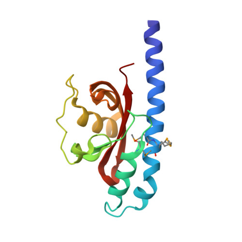
Structure and Proposed Mechanism for the pH-Sensing Helicobacter pylori Chemoreceptor TlpB.

Goers Sweeney, E.Henderson, J.N.Goers, J.Wreden, C.Hicks, K.G.Foster, J.K.Parthasarathy, R.Remington, S.J.Guillemin, K.

(2012) Structure 20: 1177-1188

  • DOI: https://doi.org/10.1016/j.str.2012.04.021
  • Primary Citation of Related Structures:  
    3UB6, 3UB7, 3UB8, 3UB9, 4EXO

  • PubMed Abstract: 

    pH sensing is crucial for survival of most organisms, yet the molecular basis of such sensing is poorly understood. Here, we present an atomic resolution structure of the periplasmic portion of the acid-sensing chemoreceptor, TlpB, from the gastric pathogen Helicobacter pylori. The structure reveals a universal signaling fold, a PAS domain, with a molecule of urea bound with high affinity. Through biophysical, biochemical, and in vivo mutagenesis studies, we show that urea and the urea-binding site residues play critical roles in the ability of H. pylori to sense acid. Our signaling model predicts that protonation events at Asp114, affected by changes in pH, dictate the stability of TlpB through urea binding.


  • Organizational Affiliation

    Institute of Molecular Biology, University of Oregon, Eugene, OR 97403, USA.


Macromolecules
Find similar proteins by:  (by identity cutoff)  |  3D Structure
Entity ID: 1
MoleculeChains Sequence LengthOrganismDetailsImage
Methyl-accepting chemotaxis protein146Vibrio parahaemolyticusMutation(s): 0 
Gene Names: GI:28896957VP0183
UniProt
Find proteins for Q87T87 (Vibrio parahaemolyticus serotype O3:K6 (strain RIMD 2210633))
Explore Q87T87 
Go to UniProtKB:  Q87T87
Entity Groups  
Sequence Clusters30% Identity50% Identity70% Identity90% Identity95% Identity100% Identity
UniProt GroupQ87T87
Sequence Annotations
Expand
  • Reference Sequence
Small Molecules
Ligands 1 Unique
IDChains Name / Formula / InChI Key2D Diagram3D Interactions
PYR
Query on PYR

Download Ideal Coordinates CCD File 
B [auth A]PYRUVIC ACID
C3 H4 O3
LCTONWCANYUPML-UHFFFAOYSA-N
Modified Residues  1 Unique
IDChains TypeFormula2D DiagramParent
MSE
Query on MSE
A
L-PEPTIDE LINKINGC5 H11 N O2 SeMET
Experimental Data & Validation

Experimental Data

  • Method: X-RAY DIFFRACTION
  • Resolution: 1.90 Å
  • R-Value Free: 0.206 
  • R-Value Work: 0.156 
  • R-Value Observed: 0.159 
  • Space Group: I 4 2 2
Unit Cell:
Length ( Å )Angle ( ˚ )
a = 112.268α = 90
b = 112.268β = 90
c = 62.731γ = 90
Software Package:
Software NamePurpose
REFMACrefinement
PDB_EXTRACTdata extraction

Structure Validation

View Full Validation Report


Entry History 

Deposition Data

Revision History  (Full details and data files)

  • Version 1.0: 2012-05-30
    Type: Initial release
  • Version 1.1: 2012-08-29
    Changes: Database references
  • Version 1.2: 2017-11-15
    Changes: Refinement description
  • Version 1.3: 2023-09-13
    Changes: Data collection, Database references, Derived calculations, Refinement description
  • Version 2.0: 2023-11-15
    Changes: Atomic model, Data collection