4EVE

Crystal Structure HP-NAP from strain YS29 in apo form


Experimental Data Snapshot

  • Method: X-RAY DIFFRACTION
  • Resolution: 2.10 Å
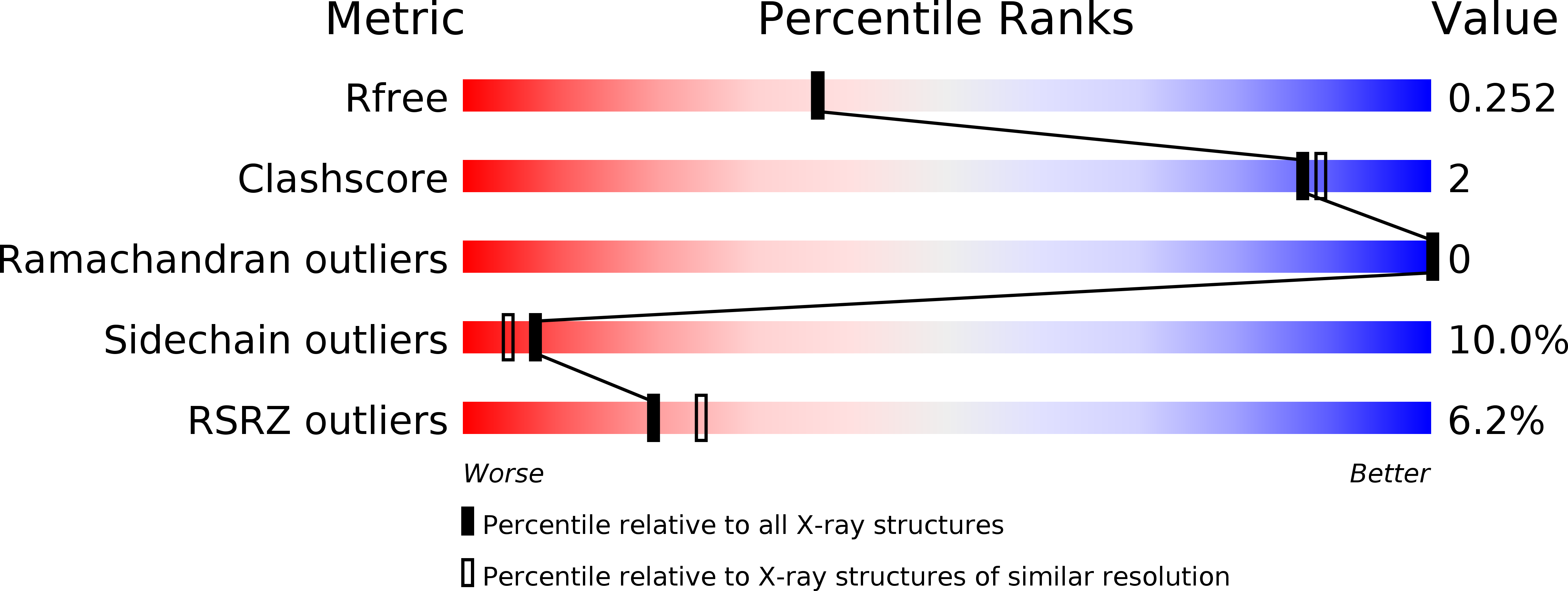
  • R-Value Free: 0.252 
  • R-Value Work: 0.216 
  • R-Value Observed: 0.220 

wwPDB Validation   3D Report Full Report


This is version 1.1 of the entry. See complete history


Literature

Crystal structure of Helicobacter pylori neutrophil-activating protein with a di-nuclear ferroxidase center in a zinc or cadmium-bound form

Yokoyama, H.Tsuruta, O.Akao, N.Fujii, S.

(2012) Biochem Biophys Res Commun 422: 745-750

  • DOI: https://doi.org/10.1016/j.bbrc.2012.05.073
  • Primary Citation of Related Structures:  
    4EVB, 4EVC, 4EVD, 4EVE

  • PubMed Abstract: 

    Helicobacter pylori neutrophil-activating protein (HP-NAP) is a Dps-like iron storage protein forming a dodecameric shell, and promotes adhesion of neutrophils to endothelial cells. The crystal structure of HP-NAP in a Zn(2+)- or Cd(2+)-bound form reveals the binding of two zinc or two cadmium ions and their bridged water molecule at the ferroxidase center (FOC). The two zinc ions are coordinated in a tetrahedral manner to the conserved residues among HP-NAP and Dps proteins. The two cadmium ions are coordinated in a trigonal-bipyramidal and distorted octahedral manner. In both structures, the second ion is more weakly coordinated than the first. Another zinc ion is found inside of the negatively-charged threefold-related pore, which is suitable for metal ions to pass through.


  • Organizational Affiliation

    School of Pharmaceutical Sciences, University of Shizuoka, 52-1 Yada, Shizuoka 422-8526, Japan. h-yokoya@u-shizuoka-ken.ac.jp


Macromolecules
Find similar proteins by:  (by identity cutoff)  |  3D Structure
Entity ID: 1
MoleculeChains Sequence LengthOrganismDetailsImage
Neutrophil-activating protein164Helicobacter pyloriMutation(s): 0 
Gene Names: napA
UniProt
Find proteins for G1UIZ2 (Helicobacter pylori)
Explore G1UIZ2 
Go to UniProtKB:  G1UIZ2
Entity Groups  
Sequence Clusters30% Identity50% Identity70% Identity90% Identity95% Identity100% Identity
UniProt GroupG1UIZ2
Sequence Annotations
Expand
  • Reference Sequence
Experimental Data & Validation

Experimental Data

  • Method: X-RAY DIFFRACTION
  • Resolution: 2.10 Å
  • R-Value Free: 0.252 
  • R-Value Work: 0.216 
  • R-Value Observed: 0.220 
  • Space Group: F 4 3 2
Unit Cell:
Length ( Å )Angle ( ˚ )
a = 187.79α = 90
b = 187.79β = 90
c = 187.79γ = 90
Software Package:
Software NamePurpose
REFMACrefinement
PDB_EXTRACTdata extraction
HKL-2000data collection
HKL-2000data reduction
HKL-2000data scaling
MOLREPphasing

Structure Validation

View Full Validation Report


Entry History 

Deposition Data

Revision History  (Full details and data files)

  • Version 1.0: 2012-06-27
    Type: Initial release
  • Version 1.1: 2023-11-08
    Changes: Data collection, Database references, Derived calculations, Refinement description