4DPR

Structure of human Leukotriene A4 hydrolase in complex with inhibitor captopril


Experimental Data Snapshot

  • Method: X-RAY DIFFRACTION
  • Resolution: 2.02 Å
  • R-Value Free: 0.228 
  • R-Value Work: 0.174 
  • R-Value Observed: 0.177 

wwPDB Validation   3D Report Full Report


Ligand Structure Quality Assessment 


This is version 1.4 of the entry. See complete history


Literature

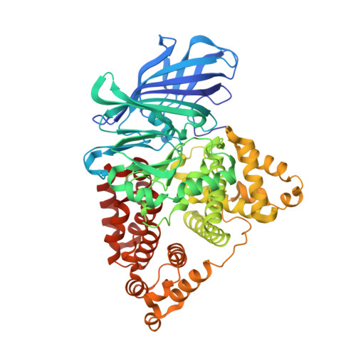
Capturing LTA4hydrolase in action: Insights to the chemistry and dynamics of chemotactic LTB4synthesis.

Stsiapanava, A.Samuelsson, B.Haeggstrom, J.Z.

(2017) Proc Natl Acad Sci U S A 114: 9689-9694

  • DOI: https://doi.org/10.1073/pnas.1710850114
  • Primary Citation of Related Structures:  
    4DPR, 5NI2, 5NI4, 5NI6, 5NIA, 5NID, 5NIE

  • PubMed Abstract: 

    Human leukotriene (LT) A 4 hydrolase/aminopeptidase (LTA 4 H) is a bifunctional enzyme that converts the highly unstable epoxide intermediate LTA 4 into LTB 4 , a potent leukocyte activating agent, while the aminopeptidase activity cleaves and inactivates the chemotactic tripeptide Pro-Gly-Pro. Here, we describe high-resolution crystal structures of LTA 4 H complexed with LTA 4 , providing the structural underpinnings of the enzyme's unique epoxide hydrolase (EH) activity, involving Zn 2+ , Y383, E271, D375, and two catalytic waters. The structures reveal that a single catalytic water is involved in both catalytic activities of LTA 4 H, alternating between epoxide ring opening and peptide bond hydrolysis, assisted by E271 and E296, respectively. Moreover, we have found two conformations of LTA 4 H, uncovering significant domain movements. The resulting structural alterations indicate that LTA 4 entrance into the active site is a dynamic process that includes rearrangement of three moving domains to provide fast and efficient alignment and processing of the substrate. Thus, the movement of one dynamic domain widens the active site entrance, while another domain acts like a lid, opening and closing access to the hydrophobic tunnel, which accommodates the aliphatic tale of LTA 4 during EH reaction. The enzyme-LTA 4 complex structures and dynamic domain movements provide critical insights for development of drugs targeting LTA 4 H.


  • Organizational Affiliation

    Division of Physiological Chemistry II, Department of Medical Biochemistry and Biophysics, Karolinska Institutet, S-171 77 Stockholm, Sweden.


Macromolecules
Find similar proteins by:  (by identity cutoff)  |  3D Structure
Entity ID: 1
MoleculeChains Sequence LengthOrganismDetailsImage
Leukotriene A-4 hydrolase611Homo sapiensMutation(s): 0 
Gene Names: LTA4HLTA4
EC: 3.3.2.6
UniProt & NIH Common Fund Data Resources
Find proteins for P09960 (Homo sapiens)
Explore P09960 
Go to UniProtKB:  P09960
PHAROS:  P09960
GTEx:  ENSG00000111144 
Entity Groups  
Sequence Clusters30% Identity50% Identity70% Identity90% Identity95% Identity100% Identity
UniProt GroupP09960
Sequence Annotations
Expand
  • Reference Sequence
Small Molecules
Ligands 5 Unique
IDChains Name / Formula / InChI Key2D Diagram3D Interactions
X8Z
Query on X8Z

Download Ideal Coordinates CCD File 
C [auth A]L-CAPTOPRIL
C9 H15 N O3 S
FAKRSMQSSFJEIM-RQJHMYQMSA-N
YB
Query on YB

Download Ideal Coordinates CCD File 
D [auth A],
E [auth A],
F [auth A],
G [auth A],
H [auth A]
YTTERBIUM (III) ION
Yb
AWSFICBXMUKWSK-UHFFFAOYSA-N
GOL
Query on GOL

Download Ideal Coordinates CCD File 
J [auth A],
K [auth A]
GLYCEROL
C3 H8 O3
PEDCQBHIVMGVHV-UHFFFAOYSA-N
ZN
Query on ZN

Download Ideal Coordinates CCD File 
B [auth A]ZINC ION
Zn
PTFCDOFLOPIGGS-UHFFFAOYSA-N
ACY
Query on ACY

Download Ideal Coordinates CCD File 
I [auth A]ACETIC ACID
C2 H4 O2
QTBSBXVTEAMEQO-UHFFFAOYSA-N
Binding Affinity Annotations 
IDSourceBinding Affinity
X8Z BindingDB:  4DPR Ki: 3700 (nM) from 1 assay(s)
Kd: min: 6250, max: 8470 (nM) from 3 assay(s)
IC50: min: 70, max: 6.30e+5 (nM) from 5 assay(s)
Experimental Data & Validation

Experimental Data

  • Method: X-RAY DIFFRACTION
  • Resolution: 2.02 Å
  • R-Value Free: 0.228 
  • R-Value Work: 0.174 
  • R-Value Observed: 0.177 
  • Space Group: P 21 21 21
Unit Cell:
Length ( Å )Angle ( ˚ )
a = 77.552α = 90
b = 87.398β = 90
c = 99.592γ = 90
Software Package:
Software NamePurpose
REFMACrefinement
PDB_EXTRACTdata extraction
MAR345data collection
XDSdata reduction
SCALAdata scaling
MOLREPphasing

Structure Validation

View Full Validation Report


Ligand Structure Quality Assessment 


Entry History 

Deposition Data

Revision History  (Full details and data files)

  • Version 1.0: 2013-02-20
    Type: Initial release
  • Version 1.1: 2017-08-23
    Changes: Database references
  • Version 1.2: 2017-11-15
    Changes: Refinement description
  • Version 1.3: 2018-04-25
    Changes: Data collection, Database references
  • Version 1.4: 2023-09-13
    Changes: Data collection, Database references, Derived calculations, Refinement description