4DKQ

Crystal structure of clade A/E 93TH057 HIV-1 gp120 core in complex with DMJ-I-228


Experimental Data Snapshot

  • Method: X-RAY DIFFRACTION
  • Resolution: 1.89 Å
  • R-Value Free: 0.233 
  • R-Value Work: 0.192 
  • R-Value Observed: 0.194 

wwPDB Validation   3D Report Full Report


Ligand Structure Quality Assessment 


This is version 1.7 of the entry. See complete history


Literature

Structure-Based Design, Synthesis, and Characterization of Dual Hotspot Small-Molecule HIV-1 Entry Inhibitors.

Lalonde, J.M.Kwon, Y.D.Jones, D.M.Sun, A.W.Courter, J.R.Soeta, T.Kobayashi, T.Princiotto, A.M.Wu, X.Schon, A.Freire, E.Kwong, P.D.Mascola, J.R.Sodroski, J.Madani, N.Smith, A.B.

(2012) J Med Chem 55: 4382-4396

  • DOI: https://doi.org/10.1021/jm300265j
  • Primary Citation of Related Structures:  
    4DKO, 4DKP, 4DKQ, 4DKR

  • PubMed Abstract: 

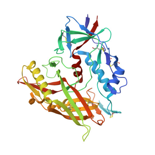
    Cellular infection by HIV-1 is initiated with a binding event between the viral envelope glycoprotein gp120 and the cellular receptor protein CD4. The CD4-gp120 interface is dominated by two hotspots: a hydrophobic gp120 cavity capped by Phe43(CD4) and an electrostatic interaction between residues Arg59(CD4) and Asp368(gp120). The CD4 mimetic small-molecule NBD-556 (1) binds within the gp120 cavity; however, 1 and related congeners demonstrate limited viral neutralization breadth. Herein, we report the design, synthesis, characterization, and X-ray structures of gp120 in complex with small molecules that simultaneously engage both binding hotspots. The compounds specifically inhibit viral infection of 42 tier 2 clades B and C viruses and are shown to be antagonists of entry into CD4-negative cells. Dual hotspot design thus provides both a means to enhance neutralization potency of HIV-1 entry inhibitors and a novel structural paradigm for inhibiting the CD4-gp120 protein-protein interaction.


  • Organizational Affiliation

    Department of Chemistry, Bryn Mawr College, Bryn Mawr, Pennsylvania 19010, USA. jlalonde@brynmawr.edu


Macromolecules
Find similar proteins by:  (by identity cutoff)  |  3D Structure
Entity ID: 1
MoleculeChains Sequence LengthOrganismDetailsImage
clade A/E 93TH057 HIV-1 gp120 core353Human immunodeficiency virus type 1 (BRU ISOLATE)Mutation(s): 0 
Gene Names: HIV-1 Env
UniProt
Find proteins for Q0ED31 (Human immunodeficiency virus 1)
Explore Q0ED31 
Go to UniProtKB:  Q0ED31
Entity Groups  
Sequence Clusters30% Identity50% Identity70% Identity90% Identity95% Identity100% Identity
UniProt GroupQ0ED31
Sequence Annotations
Expand
  • Reference Sequence
Small Molecules
Ligands 3 Unique
IDChains Name / Formula / InChI Key2D Diagram3D Interactions
0LK
Query on 0LK

Download Ideal Coordinates CCD File 
N [auth A]N-[(1S,2S)-2-carbamimidamido-2,3-dihydro-1H-inden-1-yl]-N'-(4-chloro-3-fluorophenyl)ethanediamide
C18 H17 Cl F N5 O2
ICNIHLCRBSFIFO-GJZGRUSLSA-N
EPE
Query on EPE

Download Ideal Coordinates CCD File 
M [auth A]4-(2-HYDROXYETHYL)-1-PIPERAZINE ETHANESULFONIC ACID
C8 H18 N2 O4 S
JKMHFZQWWAIEOD-UHFFFAOYSA-N
NAG
Query on NAG

Download Ideal Coordinates CCD File 
B [auth A]
C [auth A]
D [auth A]
E [auth A]
F [auth A]
B [auth A],
C [auth A],
D [auth A],
E [auth A],
F [auth A],
G [auth A],
H [auth A],
I [auth A],
J [auth A],
K [auth A],
L [auth A]
2-acetamido-2-deoxy-beta-D-glucopyranose
C8 H15 N O6
OVRNDRQMDRJTHS-FMDGEEDCSA-N
Binding Affinity Annotations 
IDSourceBinding Affinity
0LK Binding MOAD:  4DKQ Kd: 250 (nM) from 1 assay(s)
PDBBind:  4DKQ Kd: 250 (nM) from 1 assay(s)
Experimental Data & Validation

Experimental Data

  • Method: X-RAY DIFFRACTION
  • Resolution: 1.89 Å
  • R-Value Free: 0.233 
  • R-Value Work: 0.192 
  • R-Value Observed: 0.194 
  • Space Group: P 21 21 21
Unit Cell:
Length ( Å )Angle ( ˚ )
a = 63.741α = 90
b = 67.523β = 90
c = 89.255γ = 90
Software Package:
Software NamePurpose
PHENIXrefinement
PDB_EXTRACTdata extraction
HKL-2000data collection
HKL-2000data reduction
HKL-2000data scaling
PHASESphasing
REFMACrefinement

Structure Validation

View Full Validation Report


Ligand Structure Quality Assessment 


Entry History 

Revision History  (Full details and data files)

  • Version 1.0: 2012-05-02
    Type: Initial release
  • Version 1.1: 2012-05-09
    Changes: Source and taxonomy
  • Version 1.2: 2012-06-20
    Changes: Database references
  • Version 1.3: 2019-07-17
    Changes: Data collection, Derived calculations, Refinement description
  • Version 1.4: 2020-07-29
    Type: Remediation
    Reason: Carbohydrate remediation
    Changes: Data collection, Derived calculations, Structure summary
  • Version 1.5: 2021-05-19
    Changes: Source and taxonomy, Structure summary
  • Version 1.6: 2023-09-13
    Changes: Data collection, Database references, Refinement description
  • Version 1.7: 2023-12-27
    Changes: Advisory, Derived calculations