Selectivity of Pyridone- and Diphenyl Ether-Based Inhibitors for the Yersinia Pestis Fabv Enoyl-Acp Reductase.
Neckles, C., Pschibul, A., Lai, C., Hirschbeck, M., Kuper, J., Davoodi, S., Zou, J., Liu, N., Pan, P., Shah, S., Daryaee, F., Bommineni, G.R., Lai, C., Simmerling, C., Kisker, C., Tonge, P.J.(2016) Biochemistry 55: 2992
- PubMed: 27136302
- DOI: https://doi.org/10.1021/acs.biochem.5b01301
- Primary Citation of Related Structures:
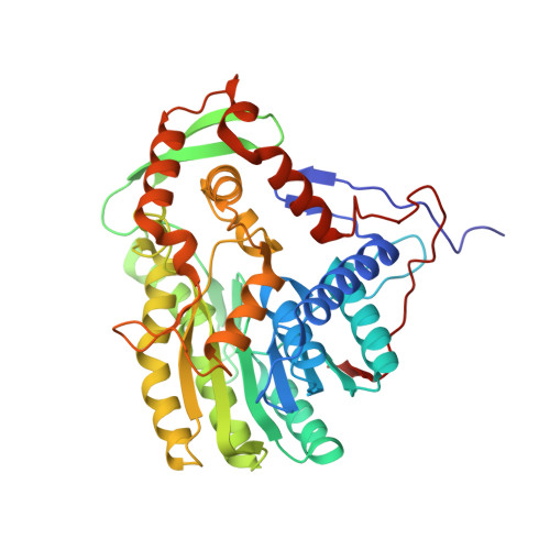
4BKQ, 4BKR, 5G2O, 5JAI, 5JAM, 5JAQ - PubMed Abstract:
The enoyl-ACP reductase (ENR) catalyzes the last reaction in the elongation cycle of the bacterial type II fatty acid biosynthesis (FAS-II) pathway. While the FabI ENR is a well-validated drug target in organisms such as Mycobacterium tuberculosis and Staphylococcus aureus, alternate ENR isoforms have been discovered in other pathogens, including the FabV enzyme that is the sole ENR in Yersinia pestis (ypFabV). Previously, we showed that the prototypical ENR inhibitor triclosan was a poor inhibitor of ypFabV and that inhibitors based on the 2-pyridone scaffold were more potent [Hirschbeck, M. (2012) Structure 20 (1), 89-100]. These studies were performed with the T276S FabV variant. In the work presented here, we describe a detailed examination of the mechanism and inhibition of wild-type ypFabV and the T276S variant. The T276S mutation significantly reduces the affinity of diphenyl ether inhibitors for ypFabV (20-fold → 100-fold). In addition, while T276S ypFabV generally displays an affinity for 2-pyridone inhibitors higher than that of the wild-type enzyme, the 4-pyridone scaffold yields compounds with similar affinity for both wild-type and T276S ypFabV. T276 is located at the N-terminus of the helical substrate-binding loop, and structural studies coupled with site-directed mutagenesis reveal that alterations in this residue modulate the size of the active site portal. Subsequently, we were able to probe the mechanism of time-dependent inhibition in this enzyme family by extending the inhibition studies to include P142W ypFabV, a mutation that results in a gain of slow-onset inhibition for the 4-pyridone PT156.
Organizational Affiliation:
Rudolf Virchow Center for Experimental Biomedicine, Institute for Structural Biology, University of Würzburg , D-97080 Würzburg, Germany.