4BEB

MUTANT (K220E) OF THE HSDR SUBUNIT OF THE ECOR124I RESTRICTION ENZYME IN COMPLEX WITH ATP


Experimental Data Snapshot

  • Method: X-RAY DIFFRACTION
  • Resolution: 2.99 Å
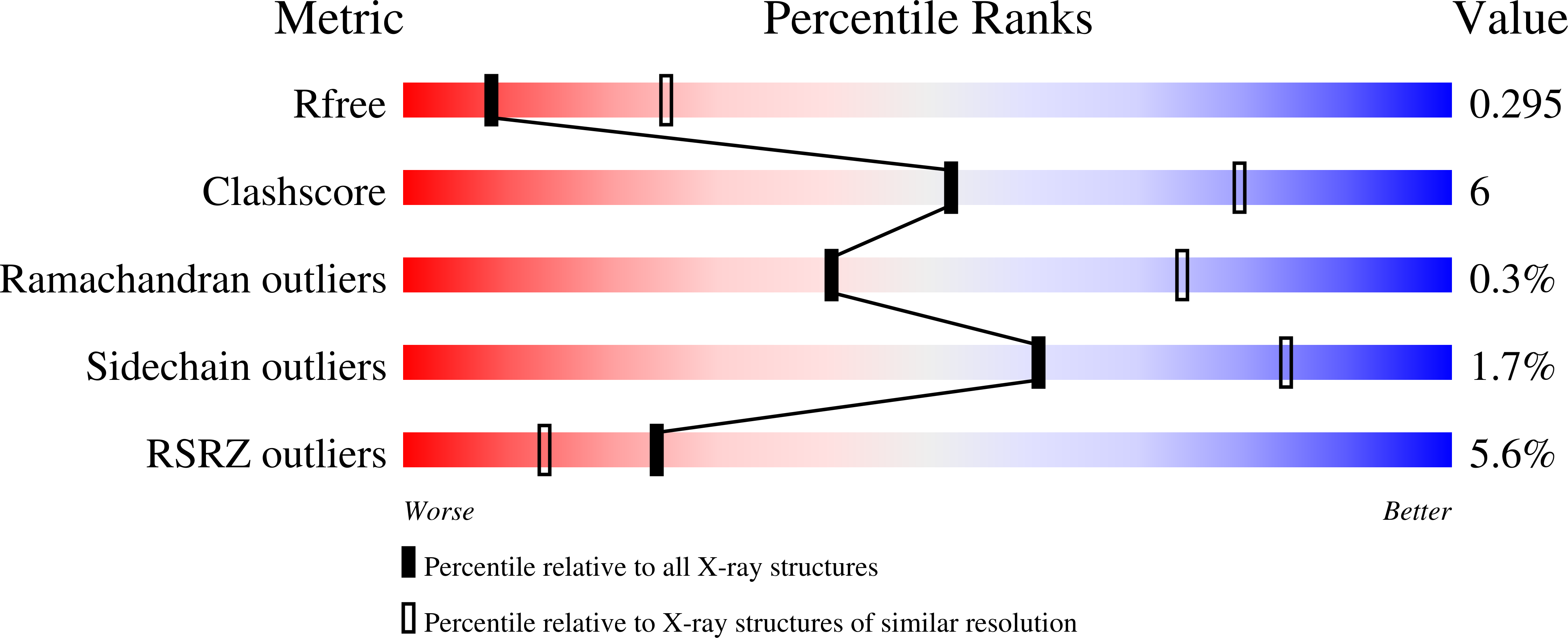
  • R-Value Free: 0.296 
  • R-Value Work: 0.255 
  • R-Value Observed: 0.256 

wwPDB Validation   3D Report Full Report


Ligand Structure Quality Assessment 


This is version 1.5 of the entry. See complete history


Literature

Functional Coupling of Duplex Translocation to DNA Cleavage in a Type I Restriction Enzyme.

Csefalvay, E.Lapkouski, M.Guzanova, A.Csefalvay, L.Baikova, T.Shevelev, I.Bialevich, V.Shamayeva, K.Janscak, P.Kuta Smatanova, I.Panjikar, S.Carey, J.Weiserova, M.Ettrich, R.

(2015) PLoS One 10: 28700

  • DOI: https://doi.org/10.1371/journal.pone.0128700
  • Primary Citation of Related Structures:  
    4BE7, 4BEB, 4BEC

  • PubMed Abstract: 

    Type I restriction-modification enzymes are multifunctional heteromeric complexes with DNA cleavage and ATP-dependent DNA translocation activities located on motor subunit HsdR. Functional coupling of DNA cleavage and translocation is a hallmark of the Type I restriction systems that is consistent with their proposed role in horizontal gene transfer. DNA cleavage occurs at nonspecific sites distant from the cognate recognition sequence, apparently triggered by stalled translocation. The X-ray crystal structure of the complete HsdR subunit from E. coli plasmid R124 suggested that the triggering mechanism involves interdomain contacts mediated by ATP. In the present work, in vivo and in vitro activity assays and crystal structures of three mutants of EcoR124I HsdR designed to probe this mechanism are reported. The results indicate that interdomain engagement via ATP is indeed responsible for signal transmission between the endonuclease and helicase domains of the motor subunit. A previously identified sequence motif that is shared by the RecB nucleases and some Type I endonucleases is implicated in signaling.


  • Organizational Affiliation

    Center for Nanobiology and Structural Biology, Institute of Microbiology and Global Change Research Center, Academy of Sciences of the Czech Republic, Zamek 136, CZ-373 33 Nove Hrady, Czech Republic.


Macromolecules
Find similar proteins by:  (by identity cutoff)  |  3D Structure
Entity ID: 1
MoleculeChains Sequence LengthOrganismDetailsImage
TYPE I RESTRICTION ENZYME HSDR
A, B, C, D
1,038Escherichia coliMutation(s): 1 
EC: 3.1.21.3
UniProt
Find proteins for P10486 (Escherichia coli)
Explore P10486 
Go to UniProtKB:  P10486
Entity Groups  
Sequence Clusters30% Identity50% Identity70% Identity90% Identity95% Identity100% Identity
UniProt GroupP10486
Sequence Annotations
Expand
  • Reference Sequence
Experimental Data & Validation

Experimental Data

  • Method: X-RAY DIFFRACTION
  • Resolution: 2.99 Å
  • R-Value Free: 0.296 
  • R-Value Work: 0.255 
  • R-Value Observed: 0.256 
  • Space Group: P 1 21 1
Unit Cell:
Length ( Å )Angle ( ˚ )
a = 127.112α = 90
b = 123.112β = 111.48
c = 160.11γ = 90
Software Package:
Software NamePurpose
PHENIXrefinement
XDSdata reduction
XSCALEdata scaling
MOLREPphasing

Structure Validation

View Full Validation Report


Ligand Structure Quality Assessment 


Entry History 

Deposition Data

Revision History  (Full details and data files)

  • Version 1.0: 2014-03-26
    Type: Initial release
  • Version 1.1: 2015-06-17
    Changes: Database references
  • Version 1.2: 2019-05-08
    Changes: Data collection, Experimental preparation, Other
  • Version 1.3: 2019-05-15
    Changes: Data collection, Experimental preparation
  • Version 1.4: 2019-07-17
    Changes: Data collection
  • Version 1.5: 2023-12-20
    Changes: Data collection, Database references, Derived calculations, Other, Refinement description