An Engineered Pii Protein Variant that Senses a Novel Ligand: Atomic Resolution Structure of the Complex with Citrate.
Zeth, K., Fokina, O., Forchhammer, K.(2012) Acta Crystallogr D Biol Crystallogr 68: 901
- PubMed: 22868755
- DOI: https://doi.org/10.1107/S0907444912016447
- Primary Citation of Related Structures:
4AFF - PubMed Abstract:
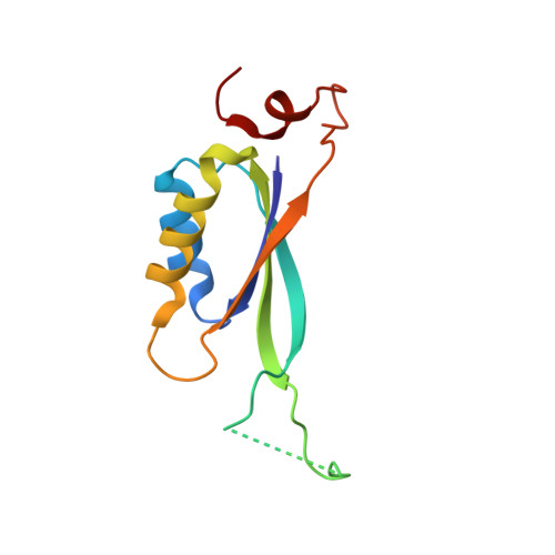
PII proteins are central signal processing units for the regulation of nitrogen metabolism in bacteria, archaea and plants. They act in response to cellular energy, carbon and nitrogen availability. The central metabolites ATP, ADP and 2-oxoglutarate, which indicate cellular energy and carbon/nitrogen abundance, bind in a highly organized manner to PII and induce effector-molecule-dependent conformational states of the T-loop. Depending on these states, PII proteins bind and modulate the activity of various regulatory targets. A mutant variant of the Synechococcus elongatus PII protein (PII-I86N) has been identified to have impaired 2-oxoglutarate binding. Here, the PII-I86N variant was cocrystallized in the presence of ATP, magnesium and citrate and its structure was solved at a resolution of 1.05 Å. The PII-I86N variant bound citrate in place of 2-oxoglutarate. Citrate binding is mediated primarily by interactions with the ATP-coordinated magnesium ion and the backbone atoms of the T-loop. Citrate binding rearranges the conformation of the T-loop and, consistent with this, citrate suppresses the binding of PII-I86N to an NAG kinase variant, which is similar to the suppression of PII-NAG kinase complex formation by 2-OG. Based on the structures of 2-OG and citrate, homocitrate was suggested as a third ligand and an efficient response towards this molecule with different functional properties was observed. Together, these data provide a first glimpse of a genetically engineered PII variant that senses a new effector molecule.
Organizational Affiliation:
Department of Protein Evolution, Max Planck Institute for Developmental Biology, Spemannstrasse 35, 72076 Tübingen, Germany. kornelius.zeth@googlemail.com