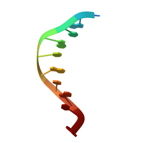
Structure of the oligonucleotide d(CGTATATACG) as a site-specific complex with nickel ions.
Abrescia, N.G., Malinina, L., Fernandez, L.G., Huynh-Dinh, T., Neidle, S., Subirana, J.A.(1999) Nucleic Acids Res 27: 1593-1599
- PubMed: 10075989
- DOI: https://doi.org/10.1093/nar/27.7.1593
- Primary Citation of Related Structures:
446D - PubMed Abstract:
In this paper we explore the application of Ni2+to the crystallization of oligonucleotides. We have determined in this way the structure of a fully alternating (Y-R) decanucleotide d(CGTATATACG) by single crystal X-ray diffraction. This is the first oligonucleotide crystal structure with an alternating 5'-(TA)3-3' central part. Alternating oligonucleotides have a particular interest since they often have a unique structure. In this case the general conformation is B-like with an alternating twist and an end-to-end interaction which involves terminal guanines. The crystal belongs to space group P41212 with a = b = 52.46, c = 101.49 A. This packing imposes a 90 degrees crossing of the symmetry related helices. This is a new way of packing for decamers. The oligonucleotide structure is characterized by the specific association with seven nickel ions, involving the N7 atom of every guanine. One of the Ni2+ions is shared between two guanines of symmetry related molecules. Until now no oligonucleotide has been crystallized in the presence of this metal ion. A novel C.A.T triplet structure has also been tentatively identified.
Organizational Affiliation:
Departament d'Enginyeria Química, Universitat Politècnica de Catalunya, Diagonal 647, Barcelona E-08028, Spain.