3ZXE

Crystal structure of Human Galectin-7 in complex with a galactose- benzylphosphate inhibitor


Experimental Data Snapshot

  • Method: X-RAY DIFFRACTION
  • Resolution: 1.67 Å
  • R-Value Free: 0.236 
  • R-Value Work: 0.205 
  • R-Value Observed: 0.206 

wwPDB Validation   3D Report Full Report


Ligand Structure Quality Assessment 


This is version 1.2 of the entry. See complete history


Literature

Inhibition Mechanism of Human Galectin-7 by a Novel Galactose-Benzylphosphate Inhibitor.

Masuyer, G.Jabeen, T.Oberg, C.T.Leffler, H.Nilsson, U.J.Acharya, K.R.

(2012) FEBS J 279: 193

  • DOI: https://doi.org/10.1111/j.1742-4658.2011.08414.x
  • Primary Citation of Related Structures:  
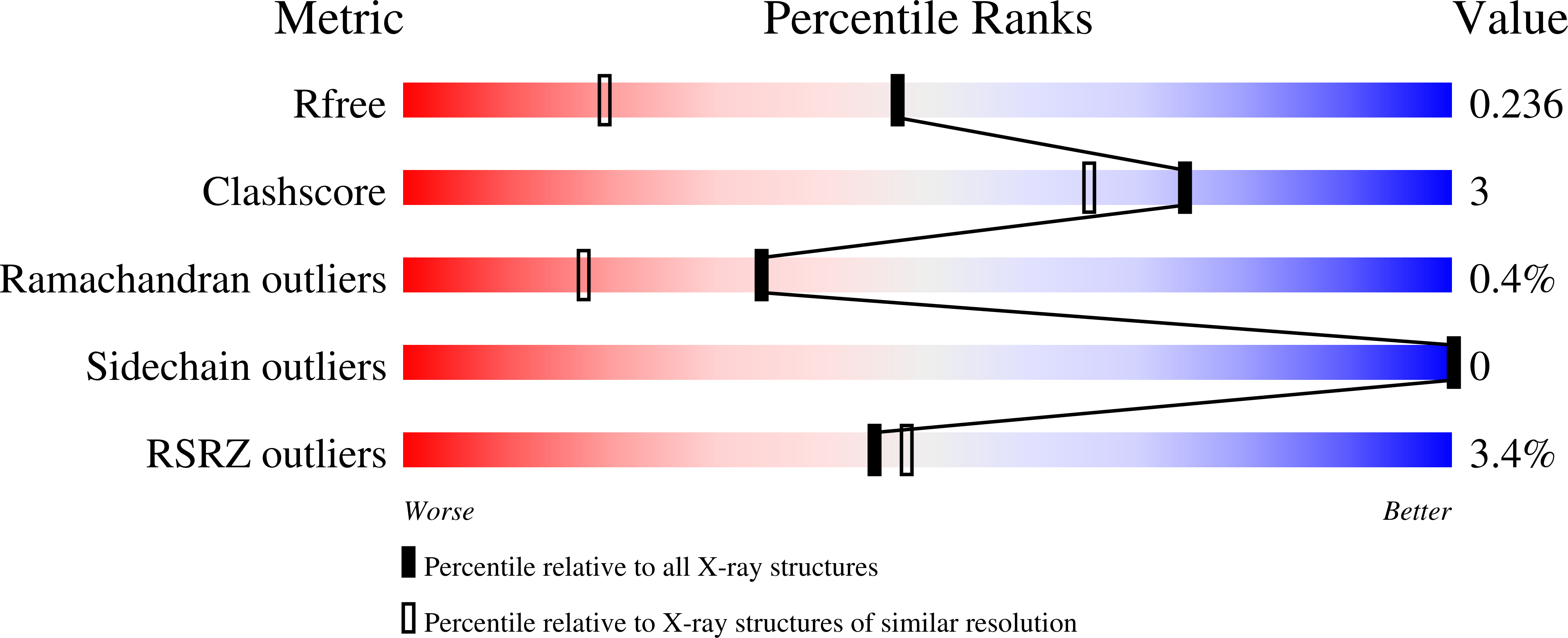
    3ZXE, 3ZXF

  • PubMed Abstract: 

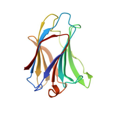
    Galectins are involved in many cellular processes due to their ability to bind carbohydrates. Understanding their functions has shown the necessity for potent and specific galectin inhibitors. Human galectin-7 (hGal-7), in particular, has been highlighted as an important marker in many types of cancer by either inhibiting or promoting tumour growth. Producing ligands able to selectively target hGal-7 will offer promising tools for deciphering cancer processes in which hGal-7 is involved as well as present potential solutions for future therapeutics. Here we report the high resolution crystal structure of hGal-7 in complex with a synthetic 2-O-benzylphosphate-galactoside inhibitor (which is > 60-fold more potent than its parent galactoside). The high resolution crystallographic analysis highlights the validity of using saccharide derivatives, conserving properties of the galactose binding, while enhanced affinity and specificity is provided by the added phosphate group. This structural information will allow the design of further inhibitors with improved potency and specificity.


  • Organizational Affiliation

    Department of Biology and Biochemistry, University of Bath, Bath, UK.


Macromolecules
Find similar proteins by:  (by identity cutoff)  |  3D Structure
Entity ID: 1
MoleculeChains Sequence LengthOrganismDetailsImage
GALECTIN-7
A, B
133Homo sapiensMutation(s): 0 
UniProt & NIH Common Fund Data Resources
Find proteins for P47929 (Homo sapiens)
Explore P47929 
Go to UniProtKB:  P47929
PHAROS:  P47929
Entity Groups  
Sequence Clusters30% Identity50% Identity70% Identity90% Identity95% Identity100% Identity
UniProt GroupP47929
Sequence Annotations
Expand
  • Reference Sequence
Small Molecules
Ligands 1 Unique
IDChains Name / Formula / InChI Key2D Diagram3D Interactions
PGZ
Query on PGZ

Download Ideal Coordinates CCD File 
C [auth A],
D [auth B]
METHYL 2-O-[(S)-(BENZYLOXY)(HYDROXY)PHOSPHORYL]-3-DEOXY-3-{[(4-METHYLPHENYL)CARBONYL]AMINO}-1-THIO-BETA-D-GALACTOPYRANOSIDE
C22 H28 N O8 P S
IDAWZVIHHBVHNS-QWPXSNKBSA-N
Binding Affinity Annotations 
IDSourceBinding Affinity
PGZ PDBBind:  3ZXE Kd: 4.50e+5 (nM) from 1 assay(s)
Binding MOAD:  3ZXE Kd: 4.50e+5 (nM) from 1 assay(s)
Experimental Data & Validation

Experimental Data

  • Method: X-RAY DIFFRACTION
  • Resolution: 1.67 Å
  • R-Value Free: 0.236 
  • R-Value Work: 0.205 
  • R-Value Observed: 0.206 
  • Space Group: P 21 21 21
Unit Cell:
Length ( Å )Angle ( ˚ )
a = 35.287α = 90
b = 53.459β = 90
c = 138.646γ = 90
Software Package:
Software NamePurpose
REFMACrefinement
HKL-2000data reduction
HKL-2000data scaling
PHASERphasing

Structure Validation

View Full Validation Report


Ligand Structure Quality Assessment 


Entry History 

Deposition Data

Revision History  (Full details and data files)

  • Version 1.0: 2011-11-30
    Type: Initial release
  • Version 1.1: 2012-01-25
    Changes: Other
  • Version 1.2: 2023-12-20
    Changes: Data collection, Database references, Derived calculations, Other, Refinement description