3ZH0

Functional and structural role of the N-terminal extension in Methanosarcina acetivorans protoglobin


Experimental Data Snapshot

  • Method: X-RAY DIFFRACTION
  • Resolution: 2.00 Å
  • R-Value Free: 0.260 
  • R-Value Work: 0.199 
  • R-Value Observed: 0.202 

wwPDB Validation   3D Report Full Report


This is version 1.2 of the entry. See complete history


Literature

Functional and Structural Role of the N-Terminal Extension in Methanosarcina Acetivorans Protoglobin.

Ciaccio, C.Pesce, A.Tundo, G.R.Tilleman, L.Bertolacci, L.Dewilde, S.Moens, L.Ascenzi, P.Bolognesi, M.Nardini, M.Coletta, M.

(2013) Biochim Biophys Acta 1834: 1813

  • DOI: https://doi.org/10.1016/j.bbapap.2013.02.026
  • Primary Citation of Related Structures:  
    3ZH0

  • PubMed Abstract: 

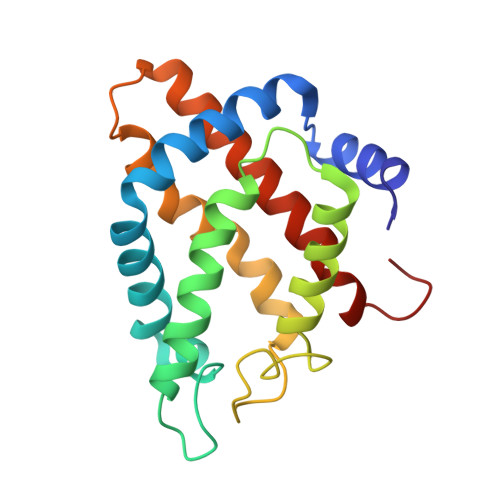
    Functional and structural properties of protoglobin from Methanosarcina acetivorans, whose Cys(101)E20 residue was mutated to Ser (MaPgb*), and of mutants missing either the first 20 N-terminal amino acids (MaPgb*-ΔN20 mutant), or the first 33 N-terminal amino acids [N-terminal loop of 20 amino acids and a 13-residue Z-helix, preceding the globin fold A-helix; (MaPgb*-ΔN20Z mutant)] have been investigated. In keeping with the MaPgb*-ΔN20 mutant crystal structure, here reported at 2.0Å resolution, which shows an increased exposure of the haem propionates to the solvent, the analysis of ligand binding kinetics highlights high accessibility of ligands to the haem pocket in ferric MaPgb*-ΔN20. CO binding to ferrous MaPgb*-ΔN20 displays a marked biphasic behavior, with a fast binding process close to that observed in MaPgb* and a slow carbonylation process, characterized by a rate-limiting step. Conversely, removal of the first 33 residues induces a substantial perturbation of the overall MaPgb* structure, with loss of α-helical content and potential partial collapse of the protein chain. As such, ligand binding kinetics are characterized by very slow rates that are independent of ligand concentration, this being indicative of a high energy barrier for ligand access to the haem, possibly due to localized misfolding. This article is part of a Special Issue entitled: Oxygen Binding and Sensing Proteins.


  • Organizational Affiliation

    Department of Clinical Sciences and Translational Medicine, University of Roma Tor Vergata, Roma, Italy.


Macromolecules
Find similar proteins by:  (by identity cutoff)  |  3D Structure
Entity ID: 1
MoleculeChains Sequence LengthOrganismDetailsImage
PROTOGLOBIN
A, B, C, D
176Methanosarcina acetivorans C2AMutation(s): 1 
UniProt
Find proteins for Q8TLY9 (Methanosarcina acetivorans (strain ATCC 35395 / DSM 2834 / JCM 12185 / C2A))
Explore Q8TLY9 
Go to UniProtKB:  Q8TLY9
Entity Groups  
Sequence Clusters30% Identity50% Identity70% Identity90% Identity95% Identity100% Identity
UniProt GroupQ8TLY9
Sequence Annotations
Expand
  • Reference Sequence
Small Molecules
Ligands 3 Unique
IDChains Name / Formula / InChI Key2D Diagram3D Interactions
HEM
Query on HEM

Download Ideal Coordinates CCD File 
E [auth A],
I [auth B],
L [auth C],
R [auth D]
PROTOPORPHYRIN IX CONTAINING FE
C34 H32 Fe N4 O4
KABFMIBPWCXCRK-RGGAHWMASA-L
GOL
Query on GOL

Download Ideal Coordinates CCD File 
G [auth A]
H [auth A]
K [auth B]
N [auth C]
O [auth C]
G [auth A],
H [auth A],
K [auth B],
N [auth C],
O [auth C],
P [auth C],
Q [auth C],
T [auth D]
GLYCEROL
C3 H8 O3
PEDCQBHIVMGVHV-UHFFFAOYSA-N
FMT
Query on FMT

Download Ideal Coordinates CCD File 
F [auth A],
J [auth B],
M [auth C],
S [auth D]
FORMIC ACID
C H2 O2
BDAGIHXWWSANSR-UHFFFAOYSA-N
Experimental Data & Validation

Experimental Data

  • Method: X-RAY DIFFRACTION
  • Resolution: 2.00 Å
  • R-Value Free: 0.260 
  • R-Value Work: 0.199 
  • R-Value Observed: 0.202 
  • Space Group: P 1 21 1
Unit Cell:
Length ( Å )Angle ( ˚ )
a = 80.67α = 90
b = 48.62β = 92.61
c = 92.03γ = 90
Software Package:
Software NamePurpose
REFMACrefinement
MOSFLMdata reduction
SCALAdata scaling
PHASERphasing

Structure Validation

View Full Validation Report


Entry History 

Deposition Data

Revision History  (Full details and data files)

  • Version 1.0: 2013-03-27
    Type: Initial release
  • Version 1.1: 2013-08-07
    Changes: Database references
  • Version 1.2: 2023-12-20
    Changes: Data collection, Database references, Derived calculations, Other, Refinement description