3ZBF

Structure of Human ROS1 Kinase Domain in Complex with Crizotinib


Experimental Data Snapshot

  • Method: X-RAY DIFFRACTION
  • Resolution: 2.20 Å
  • R-Value Free: 0.282 
  • R-Value Work: 0.233 
  • R-Value Observed: 0.236 

wwPDB Validation   3D Report Full Report


Ligand Structure Quality Assessment 


This is version 1.4 of the entry. See complete history


Literature

Acquired Resistance to Crizotinib from a Mutation in Cd74-Ros1

Awad, M.M.Katayama, R.Mctigue, M.Liu, W.Deng, Y.Brooun, A.Friboulet, L.Huang, D.Falk, M.D.Timofeevski, S.Wilner, K.D.Lockerman, E.L.Khan, T.M.Mahmood, S.Gainor, J.F.Digumarthy, S.R.Stone, J.R.Mino-Kenudson, M.Christensen, J.G.Iafrate, J.Engelman, J.A.Shaw, A.T.

(2013) N Engl J Med 368: 2395

  • DOI: https://doi.org/10.1056/NEJMoa1215530
  • Primary Citation of Related Structures:  
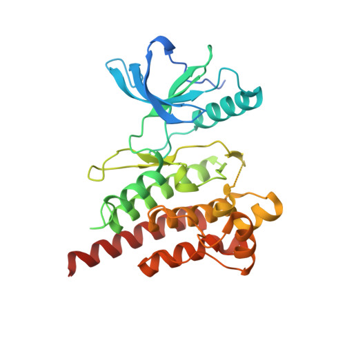
    3ZBF

  • PubMed Abstract: 

    Crizotinib, an inhibitor of anaplastic lymphoma kinase (ALK), has also recently shown efficacy in the treatment of lung cancers with ROS1 translocations. Resistance to crizotinib developed in a patient with metastatic lung adenocarcinoma harboring a CD74-ROS1 rearrangement who had initially shown a dramatic response to treatment. We performed a biopsy of a resistant tumor and identified an acquired mutation leading to a glycine-to-arginine substitution at codon 2032 in the ROS1 kinase domain. Although this mutation does not lie at the gatekeeper residue, it confers resistance to ROS1 kinase inhibition through steric interference with drug binding. The same resistance mutation was observed at all the metastatic sites that were examined at autopsy, suggesting that this mutation was an early event in the clonal evolution of resistance. (Funded by Pfizer and others; ClinicalTrials.gov number, NCT00585195.).


  • Organizational Affiliation

    Department of Medicine, Massachusetts General Hospital Cancer Center, Boston, MA 02114, USA.


Macromolecules
Find similar proteins by:  (by identity cutoff)  |  3D Structure
Entity ID: 1
MoleculeChains Sequence LengthOrganismDetailsImage
PROTO-ONCOGENE TYROSINE-PROTEIN KINASE ROS322Homo sapiensMutation(s): 0 
EC: 2.7.10.1
UniProt & NIH Common Fund Data Resources
Find proteins for P08922 (Homo sapiens)
Explore P08922 
Go to UniProtKB:  P08922
PHAROS:  P08922
GTEx:  ENSG00000047936 
Entity Groups  
Sequence Clusters30% Identity50% Identity70% Identity90% Identity95% Identity100% Identity
UniProt GroupP08922
Sequence Annotations
Expand
  • Reference Sequence
Small Molecules
Ligands 1 Unique
IDChains Name / Formula / InChI Key2D Diagram3D Interactions
VGH
Query on VGH

Download Ideal Coordinates CCD File 
B [auth A]3-[(1R)-1-(2,6-dichloro-3-fluorophenyl)ethoxy]-5-(1-piperidin-4-yl-1H-pyrazol-4-yl)pyridin-2-amine
C21 H22 Cl2 F N5 O
KTEIFNKAUNYNJU-GFCCVEGCSA-N
Binding Affinity Annotations 
IDSourceBinding Affinity
VGH BindingDB:  3ZBF Ki: min: 0.6, max: 330 (nM) from 3 assay(s)
Kd: 4.1 (nM) from 1 assay(s)
IC50: min: 0.7, max: 6700 (nM) from 8 assay(s)
Binding MOAD:  3ZBF Ki: 2.1 (nM) from 1 assay(s)
PDBBind:  3ZBF IC50: 30 (nM) from 1 assay(s)
Experimental Data & Validation

Experimental Data

  • Method: X-RAY DIFFRACTION
  • Resolution: 2.20 Å
  • R-Value Free: 0.282 
  • R-Value Work: 0.233 
  • R-Value Observed: 0.236 
  • Space Group: P 64
Unit Cell:
Length ( Å )Angle ( ˚ )
a = 106.854α = 90
b = 106.854β = 90
c = 50.357γ = 120
Software Package:
Software NamePurpose
CNXrefinement
HKL-2000data reduction
SCALEPACKdata scaling
CNXphasing

Structure Validation

View Full Validation Report


Ligand Structure Quality Assessment 


Entry History 

Deposition Data

Revision History  (Full details and data files)

  • Version 1.0: 2013-06-12
    Type: Initial release
  • Version 1.1: 2013-07-03
    Changes: Database references
  • Version 1.2: 2019-04-24
    Changes: Data collection, Other, Source and taxonomy
  • Version 1.3: 2019-05-08
    Changes: Data collection, Experimental preparation
  • Version 1.4: 2023-12-20
    Changes: Data collection, Database references, Derived calculations, Refinement description