3WSU

Crystal structure of beta-mannanase from Streptomyces thermolilacinus


Experimental Data Snapshot

  • Method: X-RAY DIFFRACTION
  • Resolution: 1.60 Å
  • R-Value Free: 0.194 
  • R-Value Work: 0.160 
  • R-Value Observed: 0.161 

wwPDB Validation   3D Report Full Report


This is version 1.5 of the entry. See complete history


Literature

The loop structure of Actinomycete glycoside hydrolase family 5 mannanases governs substrate recognition

Kumagai, Y.Yamashita, K.Tagami, T.Uraji, M.Wan, K.Okuyama, M.Yao, M.Kimura, A.Hatanaka, T.

(2015) FEBS J 282: 4001-4014

  • DOI: https://doi.org/10.1111/febs.13401
  • Primary Citation of Related Structures:  
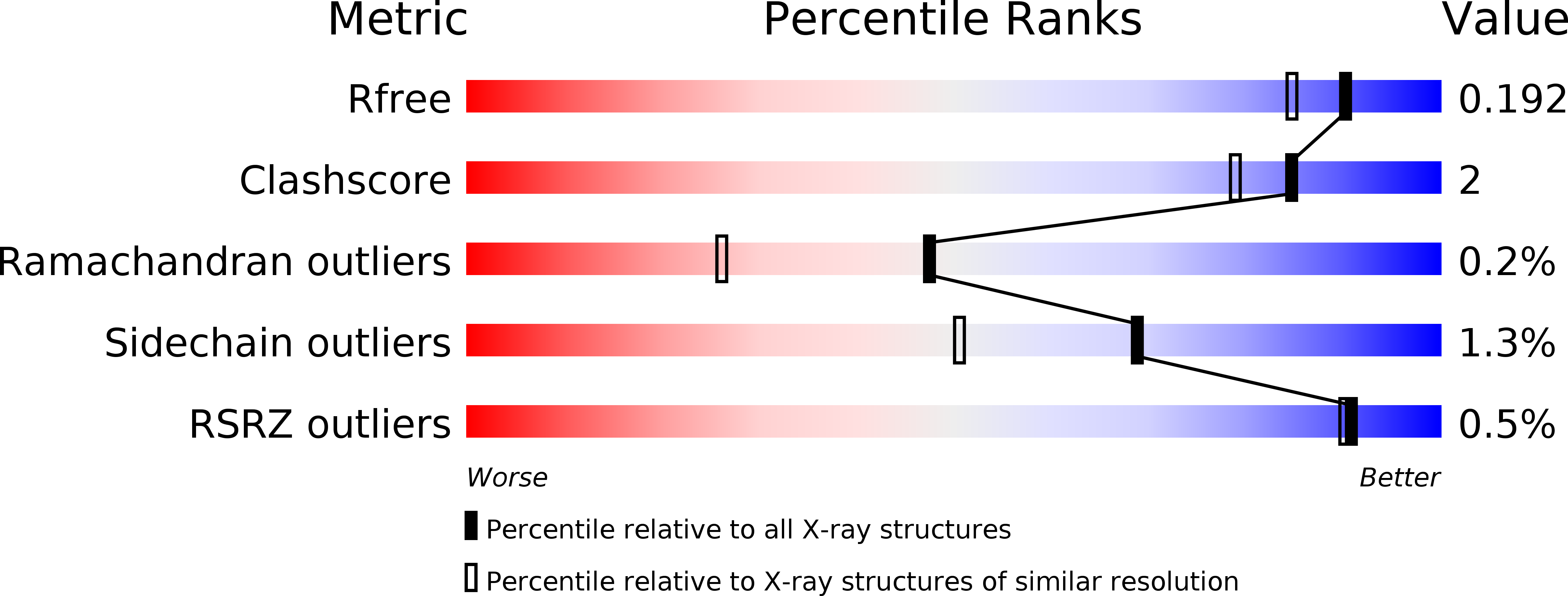
    3WSU, 4Y7E

  • PubMed Abstract: 

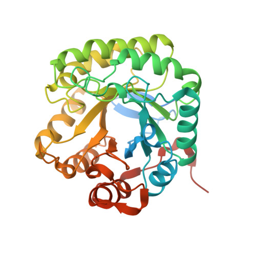
    Endo-β-1,4-mannanases from Streptomyces thermolilacinus (StMan) and Thermobifida fusca (TfMan) demonstrated different substrate specificities. StMan hydrolyzed galactosylmannooligosaccharide (GGM5; 6(III) ,6(IV) -α-d-galactosyl mannopentaose) to GGM3 and M2, whereas TfMan hydrolyzed GGM5 to GGM4 and M1. To determine the region involved in the substrate specificity, we constructed chimeric enzymes of StMan and TfMan and evaluated their substrate specificities. Moreover, the crystal structure of the catalytic domain of StMan (StMandC) and the complex structure of the inactive mutant StE273AdC with M6 were solved at resolutions of 1.60 and 1.50 Å, respectively. Structural comparisons of StMandC and the catalytic domain of TfMan lead to the identification of a subsite around -1 in StMandC that could accommodate a galactose branch. These findings demonstrate that the two loops (loop7 and loop8) are responsible for substrate recognition in GH5 actinomycete mannanases. In particular, Trp281 in loop7 of StMan, which is located in a narrow and deep cleft, plays an important role in its affinity toward linear substrates. Asp310 in loop8 of StMan specifically bound to the galactosyl unit in the -1 subsite.


  • Organizational Affiliation

    Okayama Prefectural Technology Center for Agriculture, Forestry and Fisheries, Research Institute for Biological Sciences (RIBS), Okayama, Japan.


Macromolecules
Find similar proteins by:  (by identity cutoff)  |  3D Structure
Entity ID: 1
MoleculeChains Sequence LengthOrganismDetailsImage
Beta-mannanase
A, B
346Streptomyces thermolilacinusMutation(s): 0 
Gene Names: beta-mannanase
UniProt
Find proteins for F5HR99 (Streptomyces thermolilacinus)
Explore F5HR99 
Go to UniProtKB:  F5HR99
Entity Groups  
Sequence Clusters30% Identity50% Identity70% Identity90% Identity95% Identity100% Identity
UniProt GroupF5HR99
Sequence Annotations
Expand
  • Reference Sequence
Small Molecules
Ligands 2 Unique
IDChains Name / Formula / InChI Key2D Diagram3D Interactions
GOL
Query on GOL

Download Ideal Coordinates CCD File 
E [auth A]
F [auth A]
I [auth B]
J [auth B]
K [auth B]
E [auth A],
F [auth A],
I [auth B],
J [auth B],
K [auth B],
L [auth B]
GLYCEROL
C3 H8 O3
PEDCQBHIVMGVHV-UHFFFAOYSA-N
NA
Query on NA

Download Ideal Coordinates CCD File 
C [auth A],
D [auth A],
G [auth B],
H [auth B]
SODIUM ION
Na
FKNQFGJONOIPTF-UHFFFAOYSA-N
Experimental Data & Validation

Experimental Data

  • Method: X-RAY DIFFRACTION
  • Resolution: 1.60 Å
  • R-Value Free: 0.194 
  • R-Value Work: 0.160 
  • R-Value Observed: 0.161 
  • Space Group: P 21 21 21
Unit Cell:
Length ( Å )Angle ( ˚ )
a = 65.864α = 90
b = 100.846β = 90
c = 105.244γ = 90
Software Package:
Software NamePurpose
XSCALEdata scaling
PHENIXrefinement
PDB_EXTRACTdata extraction
BSSdata collection
XDSdata reduction
XDSdata scaling
PHENIXphasing

Structure Validation

View Full Validation Report


Entry History 

Deposition Data

Revision History  (Full details and data files)

  • Version 1.0: 2015-05-20
    Type: Initial release
  • Version 1.1: 2015-09-23
    Changes: Database references
  • Version 1.2: 2015-10-14
    Changes: Other
  • Version 1.3: 2015-10-28
    Changes: Database references
  • Version 1.4: 2017-11-22
    Changes: Refinement description
  • Version 1.5: 2023-11-08
    Changes: Data collection, Database references, Derived calculations, Refinement description