3WA6

Crystal structure of tannase from Lactobacillus plantarum in the orthorhombic crystal


Experimental Data Snapshot

  • Method: X-RAY DIFFRACTION
  • Resolution: 1.80 Å
  • R-Value Free: 0.207 
  • R-Value Work: 0.178 
  • R-Value Observed: 0.178 

wwPDB Validation   3D Report Full Report


This is version 1.2 of the entry. See complete history


Literature

Crystallographic and mutational analyses of tannase from Lactobacillus plantarum

Matoba, Y.Tanaka, N.Noda, M.Higashikawa, F.Kumagai, T.Sugiyama, M.

(2013) Proteins 81: 2052-2058

  • DOI: https://doi.org/10.1002/prot.24355
  • Primary Citation of Related Structures:  
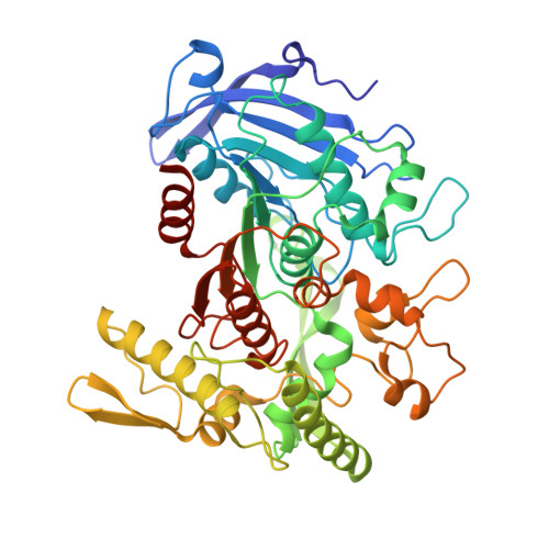
    3WA6, 3WA7

  • PubMed Abstract: 

    Tannin acylhydrolase (EC 3.1.1.20) referred commonly as tannase catalyzes the hydrolysis of the galloyl ester bond of tannins to release gallic acid. Although the enzyme is useful for various industries, the tertiary structure is not yet determined. In this study, we determined the crystal structure of tannase produced by Lactobacillus plantarum. The tannase structure belongs to a member of α/β-hydrolase superfamily with an additional "lid" domain. A glycerol molecule derived from cryoprotectant solution was accommodated into the tannase active site. The binding manner of glycerol to tannase seems to be similar to that of the galloyl moiety in the substrate.


  • Organizational Affiliation

    Department of Molecular Microbiology and Biotechnology, Graduate School of Biomedical & Health Sciences, Hiroshima University, Kasumi 1-2-3, Minami-ku, Hiroshima, 734-8551, Japan.


Macromolecules
Find similar proteins by:  (by identity cutoff)  |  3D Structure
Entity ID: 1
MoleculeChains Sequence LengthOrganismDetailsImage
tannase477Lactiplantibacillus plantarumMutation(s): 0 
EC: 3.1.1.20
UniProt
Find proteins for B3Y018 (Lactiplantibacillus plantarum)
Explore B3Y018 
Go to UniProtKB:  B3Y018
Entity Groups  
Sequence Clusters30% Identity50% Identity70% Identity90% Identity95% Identity100% Identity
UniProt GroupB3Y018
Sequence Annotations
Expand
  • Reference Sequence
Experimental Data & Validation

Experimental Data

  • Method: X-RAY DIFFRACTION
  • Resolution: 1.80 Å
  • R-Value Free: 0.207 
  • R-Value Work: 0.178 
  • R-Value Observed: 0.178 
  • Space Group: C 2 2 21
Unit Cell:
Length ( Å )Angle ( ˚ )
a = 55.87α = 90
b = 78.24β = 90
c = 200.01γ = 90
Software Package:
Software NamePurpose
HKL-2000data collection
SHARPphasing
CNSrefinement
HKL-2000data reduction
HKL-2000data scaling

Structure Validation

View Full Validation Report


Entry History 

Deposition Data

Revision History  (Full details and data files)

  • Version 1.0: 2013-07-24
    Type: Initial release
  • Version 1.1: 2013-11-06
    Changes: Database references
  • Version 1.2: 2024-03-20
    Changes: Data collection, Database references, Derived calculations