The structure of the deacetylase domain of Escherichia coli PgaB, an enzyme required for biofilm formation: a circularly permuted member of the carbohydrate esterase 4 family
Nishiyama, T., Noguchi, H., Yoshida, H., Park, S.Y., Tame, J.R.(2013) Acta Crystallogr D Biol Crystallogr 69: 44-51
- PubMed: 23275162
- DOI: https://doi.org/10.1107/S0907444912042059
- Primary Citation of Related Structures:
3VUS - PubMed Abstract:
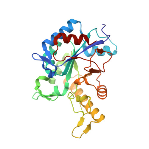
Bacterial biofilm formation is an extremely widespread phenomenon involving the secretion of a protective exopolysaccharide matrix which helps the bacteria to attach to surfaces and to overcome a variety of stresses in different environments. This matrix may also include proteins, lipids, DNA and metal ions. Its composition depends on the bacterial species and growth conditions, but one of the most widely found components is polymeric β-1,6-N-acetyl-D-glucosamine (PGA). Several studies have suggested that PGA is an essential component of biofilm and it is produced by numerous bacteria, including Escherichia coli, Staphylococcus epidermis, Yersinia pestis, Bordetella spp. and Actinobacillus spp. In E. coli, PGA production and export are dependent on four genes that form a single operon, pgaABCD, which appears to have been transferred between various species. Biofilms themselves are recognized as environments in which such horizontal gene transfer may occur. The pga operon of E. coli, which is even found in innocuous laboratory strains, is highly homologous to that from the plague bacterium Yersinia pestis, and biofilm is believed to play an important role in the transmission of Yersinia. The crystal structure of the N-terminal domain of PgaB, which has deacetylase activity, is described and compared with models of other deacetylases.
Organizational Affiliation:
Protein Design Laboratory, Graduate School of Nanobioscience, Yokohama City University, Suehiro, Kanagawa, Japan.