


Crystal Structure of 7-cyano-7-deazaguanine reductase, QueF from Vibrio cholerae complexed with NADP and PreQ0
Kim, Y., Zhang, R., Gu, M., Anderson, W.F., Joachimiak, A., CSGIDTo be published.
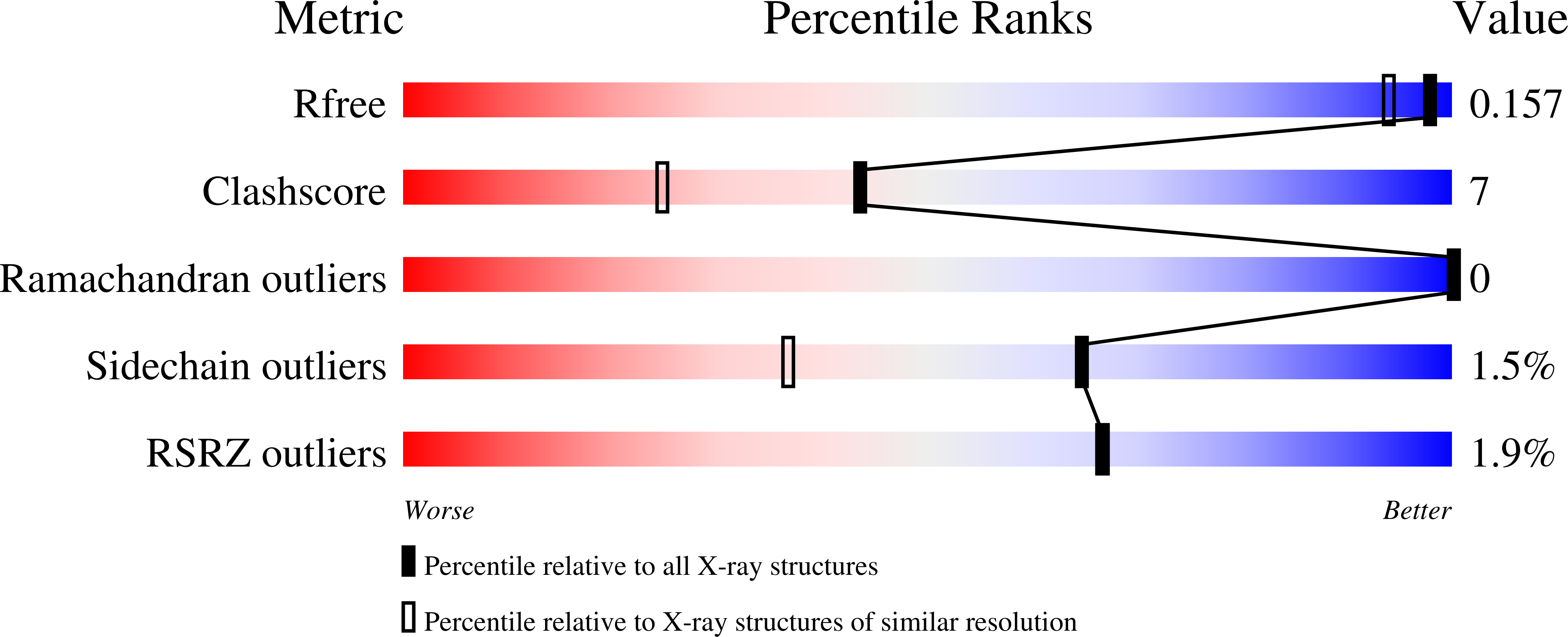
Experimental Data Snapshot
Entity ID: 1 | |||||
|---|---|---|---|---|---|
| Molecule | Chains | Sequence Length | Organism | Details | Image |
| NADPH-dependent 7-cyano-7-deazaguanine reductase | 290 | Vibrio cholerae O1 biovar El Tor str. N16961 | Mutation(s): 1 Gene Names: queF, VCM66_0859 EC: 1.7.1.13 | 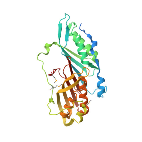 | |
UniProt | |||||
Find proteins for Q9KTK0 (Vibrio cholerae serotype O1 (strain ATCC 39315 / El Tor Inaba N16961)) Explore Q9KTK0 Go to UniProtKB: Q9KTK0 | |||||
Entity Groups | |||||
| Sequence Clusters | 30% Identity50% Identity70% Identity90% Identity95% Identity100% Identity | ||||
| UniProt Group | Q9KTK0 | ||||
Sequence AnnotationsExpand | |||||
| |||||
| Ligands 3 Unique | |||||
|---|---|---|---|---|---|
| ID | Chains | Name / Formula / InChI Key | 2D Diagram | 3D Interactions | |
| NAP Query on NAP | F [auth A], R [auth D] | NADP NICOTINAMIDE-ADENINE-DINUCLEOTIDE PHOSPHATE C21 H28 N7 O17 P3 XJLXINKUBYWONI-NNYOXOHSSA-N |  | ||
| PRF Query on PRF | E [auth A], I [auth B], O [auth C], S [auth D] | 7-DEAZA-7-AMINOMETHYL-GUANINE C7 H9 N5 O MEYMBLGOKYDGLZ-UHFFFAOYSA-N |  | ||
| EDO Query on EDO | G [auth A] H [auth A] J [auth B] K [auth B] L [auth B] | 1,2-ETHANEDIOL C2 H6 O2 LYCAIKOWRPUZTN-UHFFFAOYSA-N |  | ||
| Modified Residues 1 Unique | |||||
|---|---|---|---|---|---|
| ID | Chains | Type | Formula | 2D Diagram | Parent |
| MSE Query on MSE | A, B, C, D | L-PEPTIDE LINKING | C5 H11 N O2 Se |  | MET |
| Length ( Å ) | Angle ( ˚ ) |
|---|---|
| a = 71.387 | α = 110.01 |
| b = 71.416 | β = 119.54 |
| c = 71.358 | γ = 99.46 |
| Software Name | Purpose |
|---|---|
| SBC-Collect | data collection |
| HKL-3000 | data collection |
| HKL-3000 | phasing |
| MOLREP | phasing |
| PHENIX | refinement |
| HKL-3000 | data reduction |
| HKL-3000 | data scaling |
RCSB PDB (citation) is hosted by
RCSB PDB is a member of the

