3UMO

Crystal structure of the Phosphofructokinase-2 from Escherichia coli in complex with Potassium


Experimental Data Snapshot

  • Method: X-RAY DIFFRACTION
  • Resolution: 1.70 Å
  • R-Value Free: 0.208 
  • R-Value Work: 0.181 
  • R-Value Observed: 0.183 

wwPDB Validation   3D Report Full Report


Ligand Structure Quality Assessment 


This is version 1.2 of the entry. See complete history


Literature

A Ribokinase Family Conserved Monovalent Cation Binding Site Enhances the MgATP-induced Inhibition in E. coli Phosphofructokinase-2

Baez, M.Cabrera, R.Pereira, H.M.Blanco, A.Villalobos, P.Ramirez-Sarmiento, C.A.Caniuguir, A.Guixe, V.Garratt, R.C.Babul, J.

(2013) Biophys J 105: 185-193

  • DOI: https://doi.org/10.1016/j.bpj.2013.05.028
  • Primary Citation of Related Structures:  
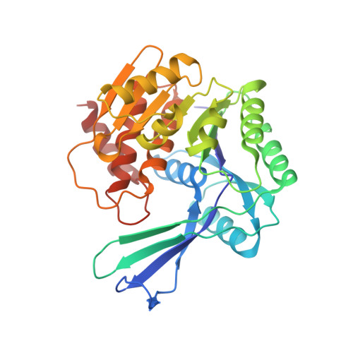
    3UMO, 3UMP

  • PubMed Abstract: 

    The presence of a regulatory site for monovalent cations that affects the conformation of the MgATP-binding pocket leading to enzyme activation has been demonstrated for ribokinases. This site is selective toward the ionic radius of the monovalent cation, accepting those larger than Na(+). Phosphofructokinase-2 (Pfk-2) from Escherichia coli is homologous to ribokinase, but unlike other ribokinase family members, presents an additional site for the nucleotide that negatively regulates its enzymatic activity. In this work, we show the effect of monovalent cations on the kinetic parameters of Pfk-2 together with its three-dimensional structure determined by x-ray diffraction in the presence of K(+) or Cs(+). Kinetic characterization of the enzyme shows that K(+) and Na(+) alter neither the kcat nor the KM values for fructose-6-P or MgATP. However, the presence of K(+) (but not Na(+)) enhances the allosteric inhibition induced by MgATP. Moreover, binding experiments show that K(+) (but not Na(+)) increases the affinity of MgATP in a saturable fashion. In agreement with the biochemical data, the crystal structure of Pfk-2 obtained in the presence of MgATP shows a cation-binding site at the conserved position predicted for the ribokinase family of proteins. This site is adjacent to the MgATP allosteric binding site and is only observed in the presence of Cs(+) or K(+). These results indicate that binding of the monovalent metal ions indirectly influences the allosteric site of Pfk-2 by increasing its affinity for MgATP with no alteration in the conformation of residues present at the catalytic site.


  • Organizational Affiliation

    Departamento de Bioquímica y Biología Molecular, Facultad de Ciencias Químicas y Farmacéuticas, Universidad de Chile, Santiago, Chile.


Macromolecules
Find similar proteins by:  (by identity cutoff)  |  3D Structure
Entity ID: 1
MoleculeChains Sequence LengthOrganismDetailsImage
6-phosphofructokinase isozyme 2
A, B
309Escherichia coli K-12Mutation(s): 0 
Gene Names: b1723JW5280PFK2pfkB
EC: 2.7.1.11
UniProt
Find proteins for P06999 (Escherichia coli (strain K12))
Explore P06999 
Go to UniProtKB:  P06999
Entity Groups  
Sequence Clusters30% Identity50% Identity70% Identity90% Identity95% Identity100% Identity
UniProt GroupP06999
Sequence Annotations
Expand
  • Reference Sequence
Small Molecules
Ligands 3 Unique
IDChains Name / Formula / InChI Key2D Diagram3D Interactions
ATP
Query on ATP

Download Ideal Coordinates CCD File 
E [auth A],
H [auth A],
I [auth B],
K [auth B]
ADENOSINE-5'-TRIPHOSPHATE
C10 H16 N5 O13 P3
ZKHQWZAMYRWXGA-KQYNXXCUSA-N
K
Query on K

Download Ideal Coordinates CCD File 
F [auth A],
L [auth B]
POTASSIUM ION
K
NPYPAHLBTDXSSS-UHFFFAOYSA-N
MG
Query on MG

Download Ideal Coordinates CCD File 
C [auth A],
D [auth A],
G [auth A],
J [auth B]
MAGNESIUM ION
Mg
JLVVSXFLKOJNIY-UHFFFAOYSA-N
Binding Affinity Annotations 
IDSourceBinding Affinity
ATP PDBBind:  3UMO Ki: 1.29e+5 (nM) from 1 assay(s)
Experimental Data & Validation

Experimental Data

  • Method: X-RAY DIFFRACTION
  • Resolution: 1.70 Å
  • R-Value Free: 0.208 
  • R-Value Work: 0.181 
  • R-Value Observed: 0.183 
  • Space Group: P 2 2 21
Unit Cell:
Length ( Å )Angle ( ˚ )
a = 43.812α = 90
b = 88.77β = 90
c = 176.12γ = 90
Software Package:
Software NamePurpose
XSCALEdata scaling
PHASERphasing
DMphasing
PHENIXrefinement
PDB_EXTRACTdata extraction
DNAdata collection
XDSdata reduction

Structure Validation

View Full Validation Report


Ligand Structure Quality Assessment 


Entry History 

Deposition Data

Revision History  (Full details and data files)

  • Version 1.0: 2012-11-14
    Type: Initial release
  • Version 1.1: 2013-07-24
    Changes: Database references
  • Version 1.2: 2023-09-13
    Changes: Data collection, Database references, Derived calculations, Refinement description