3SLE

Crystal Structure of the P107C-MauG/pre-Methylamine Dehydrogenase Complex


Experimental Data Snapshot

  • Method: X-RAY DIFFRACTION
  • Resolution: 2.52 Å
  • R-Value Free: 0.243 
  • R-Value Work: 0.174 
  • R-Value Observed: 0.178 

wwPDB Validation   3D Report Full Report


Ligand Structure Quality Assessment 


This is version 1.3 of the entry. See complete history


Literature

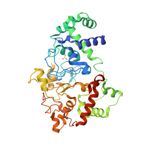
Proline 107 is a major determinant in maintaining the structure of the distal pocket and reactivity of the high-spin heme of MauG.

Feng, M.Jensen, L.M.Yukl, E.T.Wei, X.Liu, A.Wilmot, C.M.Davidson, V.L.

(2012) Biochemistry 51: 1598-1606

  • DOI: https://doi.org/10.1021/bi201882e
  • Primary Citation of Related Structures:  
    3SJL, 3SLE, 3SVW

  • PubMed Abstract: 

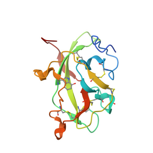
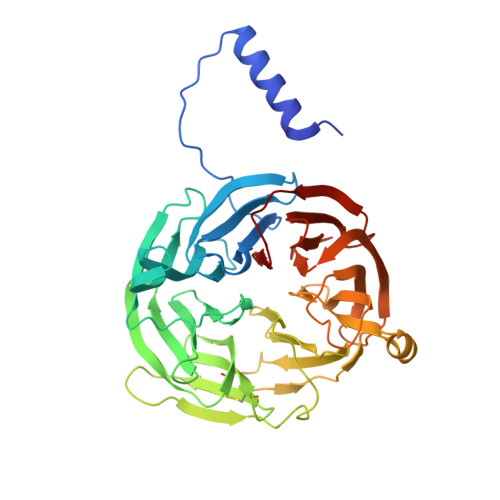
    The diheme enzyme MauG catalyzes a six-electron oxidation required for posttranslational modification of a precursor of methylamine dehydrogenase (preMADH) to complete the biosynthesis of its protein-derived tryptophan tryptophylquinone (TTQ) cofactor. Crystallographic studies had shown that Pro107, which resides in the distal pocket of the high-spin heme of MauG, changes conformation upon binding of CO or NO to the heme iron. In this study, Pro107 was converted to Cys, Val, and Ser by site-directed mutagenesis. The structures of each of these MauG mutant proteins in complex with preMADH were determined, as were their physical and catalytic properties. P107C MauG was inactive, and the crystal structure revealed that Cys107 had been oxidatively modified to a sulfinic acid. Mass spectrometry revealed that this modification was present prior to crystallization. P107V MauG exhibited spectroscopic and catalytic properties that were similar to those of wild-type MauG, but P107V MauG was more susceptible to oxidative damage. The P107S mutation caused a structural change that resulted in the five-coordinate high-spin heme being converted to a six-coordinate heme with a distal axial ligand provided by Glu113. EPR and resonance Raman spectroscopy revealed this heme remained high-spin but with greatly increased rhombicity as compared to that of the axial signal of wild-type MauG. P107S MauG was resistant to reduction by dithionite and reaction with H(2)O(2) and unable to catalyze TTQ biosynthesis. These results show that the presence of Pro107 is critical in maintaining the proper structure of the distal heme pocket of the high-spin heme of MauG, allowing exogenous ligands to bind and directing the reactivity of the heme-activated oxygen during catalysis, thus minimizing the oxidation of other residues of MauG.


  • Organizational Affiliation

    Department of Chemistry, Tougaloo College, Tougaloo, Mississippi 39174, United States.


Macromolecules
Find similar proteins by:  (by identity cutoff)  |  3D Structure
Entity ID: 1
MoleculeChains Sequence LengthOrganismDetailsImage
Methylamine utilization protein MauG
A, B
373Paracoccus denitrificans PD1222Mutation(s): 1 
Gene Names: mauGPden_4736
EC: 1
UniProt
Find proteins for Q51658 (Paracoccus denitrificans (strain Pd 1222))
Explore Q51658 
Go to UniProtKB:  Q51658
Entity Groups  
Sequence Clusters30% Identity50% Identity70% Identity90% Identity95% Identity100% Identity
UniProt GroupQ51658
Sequence Annotations
Expand
  • Reference Sequence
Find similar proteins by:  (by identity cutoff)  |  3D Structure
Entity ID: 2
MoleculeChains Sequence LengthOrganismDetailsImage
Methylamine dehydrogenase light chain
C, E
137Paracoccus denitrificans PD1222Mutation(s): 0 
Gene Names: mauA
EC: 1.4.99.3
UniProt
Find proteins for P22619 (Paracoccus denitrificans)
Explore P22619 
Go to UniProtKB:  P22619
Entity Groups  
Sequence Clusters30% Identity50% Identity70% Identity90% Identity95% Identity100% Identity
UniProt GroupP22619
Sequence Annotations
Expand
  • Reference Sequence
Find similar proteins by:  (by identity cutoff)  |  3D Structure
Entity ID: 3
MoleculeChains Sequence LengthOrganismDetailsImage
Methylamine dehydrogenase heavy chain
D, F
385Paracoccus denitrificans PD1222Mutation(s): 0 
Gene Names: mauBpden_4730
EC: 1.4.99.3
UniProt
Find proteins for A1BB97 (Paracoccus denitrificans (strain Pd 1222))
Explore A1BB97 
Go to UniProtKB:  A1BB97
Entity Groups  
Sequence Clusters30% Identity50% Identity70% Identity90% Identity95% Identity100% Identity
UniProt GroupA1BB97
Sequence Annotations
Expand
  • Reference Sequence
Small Molecules
Ligands 4 Unique
IDChains Name / Formula / InChI Key2D Diagram3D Interactions
HEC
Query on HEC

Download Ideal Coordinates CCD File 
H [auth A],
I [auth A],
K [auth B],
M [auth B]
HEME C
C34 H34 Fe N4 O4
HXQIYSLZKNYNMH-LJNAALQVSA-N
EDO
Query on EDO

Download Ideal Coordinates CCD File 
N [auth B],
P [auth D]
1,2-ETHANEDIOL
C2 H6 O2
LYCAIKOWRPUZTN-UHFFFAOYSA-N
ACT
Query on ACT

Download Ideal Coordinates CCD File 
L [auth B],
O [auth D],
Q [auth F],
R [auth F]
ACETATE ION
C2 H3 O2
QTBSBXVTEAMEQO-UHFFFAOYSA-M
CA
Query on CA

Download Ideal Coordinates CCD File 
G [auth A],
J [auth B]
CALCIUM ION
Ca
BHPQYMZQTOCNFJ-UHFFFAOYSA-N
Modified Residues  2 Unique
IDChains TypeFormula2D DiagramParent
CSD
Query on CSD
A, B
L-PEPTIDE LINKINGC3 H7 N O4 SCYS
0AF
Query on 0AF
C, E
L-PEPTIDE LINKINGC11 H12 N2 O3TRP
Experimental Data & Validation

Experimental Data

  • Method: X-RAY DIFFRACTION
  • Resolution: 2.52 Å
  • R-Value Free: 0.243 
  • R-Value Work: 0.174 
  • R-Value Observed: 0.178 
  • Space Group: P 1
Unit Cell:
Length ( Å )Angle ( ˚ )
a = 55.851α = 116.21
b = 88.53β = 91.83
c = 107.698γ = 99.35
Software Package:
Software NamePurpose
Blu-Icedata collection
REFMACrefinement
HKL-2000data reduction
HKL-2000data scaling
REFMACphasing

Structure Validation

View Full Validation Report


Ligand Structure Quality Assessment 


Entry History 

Deposition Data

Revision History  (Full details and data files)

  • Version 1.0: 2012-05-02
    Type: Initial release
  • Version 1.1: 2014-11-19
    Changes: Structure summary
  • Version 1.2: 2023-09-13
    Changes: Data collection, Database references, Derived calculations, Refinement description
  • Version 1.3: 2023-12-06
    Changes: Data collection