3S90

Human vinculin head domain Vh1 (residues 1-252) in complex with murine talin (VBS33; residues 1512-1546)


Experimental Data Snapshot

  • Method: X-RAY DIFFRACTION
  • Resolution: 1.97 Å
  • R-Value Free: 0.210 
  • R-Value Work: 0.186 
  • R-Value Observed: 0.187 

wwPDB Validation   3D Report Full Report


This is version 1.6 of the entry. See complete history


Literature

Intermolecular versus intramolecular interactions of the vinculin binding site 33 of talin.

Yogesha, S.D.Sharff, A.Bricogne, G.Izard, T.

(2011) Protein Sci 20: 1471-1476

  • DOI: https://doi.org/10.1002/pro.671
  • Primary Citation of Related Structures:  
    3S90

  • PubMed Abstract: 

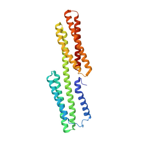
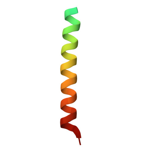
    The cytoskeletal proteins talin and vinculin are localized at cell-matrix junctions and are key regulators of cell signaling, adhesion, and migration. Talin couples integrins via its FERM domain to F-actin and is an important regulator of integrin activation and clustering. The 220 kDa talin rod domain comprises several four- and five-helix bundles that harbor amphipathic α-helical vinculin binding sites (VBSs). In its inactive state, the hydrophobic VBS residues involved in binding to vinculin are buried within these helix bundles, and the mechanical force emanating from bound integrin receptors is thought necessary for their release and binding to vinculin. The crystal structure of a four-helix bundle of talin that harbors one of these VBSs, coined VBS33, was recently determined. Here we report the crystal structure of VBS33 in complex with vinculin at 2 Å resolution. Notably, comparison of the apo and vinculin bound structures shows that intermolecular interactions of the VBS33 α-helix with vinculin are more extensive than the intramolecular interactions of the VBS33 within the talin four-helix bundle.


  • Organizational Affiliation

    Cell Adhesion Laboratory, Department of Cancer Biology, The Scripps Research Institute, Jupiter, FL 33458, USA.


Macromolecules
Find similar proteins by:  (by identity cutoff)  |  3D Structure
Entity ID: 1
MoleculeChains Sequence LengthOrganismDetailsImage
Vinculin
A, B
253Homo sapiensMutation(s): 0 
Gene Names: VCL
UniProt & NIH Common Fund Data Resources
Find proteins for P18206 (Homo sapiens)
Explore P18206 
Go to UniProtKB:  P18206
PHAROS:  P18206
GTEx:  ENSG00000035403 
Entity Groups  
Sequence Clusters30% Identity50% Identity70% Identity90% Identity95% Identity100% Identity
UniProt GroupP18206
Sequence Annotations
Expand
  • Reference Sequence
Find similar proteins by:  (by identity cutoff)  |  3D Structure
Entity ID: 2
MoleculeChains Sequence LengthOrganismDetailsImage
Talin-1
C, D
40Mus musculusMutation(s): 0 
Gene Names: Tln1Tln
UniProt
Find proteins for P26039 (Mus musculus)
Explore P26039 
Go to UniProtKB:  P26039
Entity Groups  
Sequence Clusters30% Identity50% Identity70% Identity90% Identity95% Identity100% Identity
UniProt GroupP26039
Sequence Annotations
Expand
  • Reference Sequence
Experimental Data & Validation

Experimental Data

  • Method: X-RAY DIFFRACTION
  • Resolution: 1.97 Å
  • R-Value Free: 0.210 
  • R-Value Work: 0.186 
  • R-Value Observed: 0.187 
  • Space Group: P 1
Unit Cell:
Length ( Å )Angle ( ˚ )
a = 48.83α = 83.27
b = 61.18β = 79.05
c = 71.42γ = 69.79
Software Package:
Software NamePurpose
MAR345data collection
MOLREPphasing
BUSTERrefinement
autoPROCdata scaling
XDSdata reduction
SCALAdata scaling

Structure Validation

View Full Validation Report


Entry History 

Deposition Data

Revision History  (Full details and data files)

  • Version 1.0: 2011-06-22
    Type: Initial release
  • Version 1.1: 2011-07-13
    Changes: Version format compliance
  • Version 1.2: 2012-04-25
    Changes: Database references
  • Version 1.3: 2012-12-19
    Changes: Database references
  • Version 1.4: 2014-10-29
    Changes: Structure summary
  • Version 1.5: 2017-11-08
    Changes: Refinement description
  • Version 1.6: 2023-09-13
    Changes: Data collection, Database references, Refinement description