


Flexibility and plasticity of EF-hand motifs: Structure of Calcium Binding Protein-1 from Entamoeba histolytica in complex with Pb2+, Ba2+, and Sr2+.
Kumar, S., Kumar, S., Ahmad, E., Khan, R.H., Gourinath, S.To be published.
Experimental Data Snapshot
wwPDB Validation 3D Report Full Report

Entity ID: 1 | |||||
|---|---|---|---|---|---|
| Molecule | Chains | Sequence Length | Organism | Details | Image |
| Calcium-binding protein | 134 | Entamoeba histolytica HM-1:IMSS | Mutation(s): 0 | 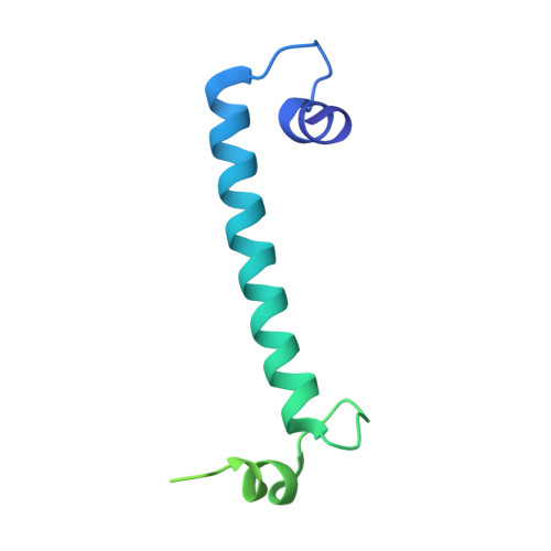 | |
UniProt | |||||
Find proteins for P38505 (Entamoeba histolytica (strain ATCC 30459 / HM-1:IMSS / ABRM)) Explore P38505 Go to UniProtKB: P38505 | |||||
Entity Groups | |||||
| Sequence Clusters | 30% Identity50% Identity70% Identity90% Identity95% Identity100% Identity | ||||
| UniProt Group | P38505 | ||||
Sequence AnnotationsExpand | |||||
| |||||
| Ligands 1 Unique | |||||
|---|---|---|---|---|---|
| ID | Chains | Name / Formula / InChI Key | 2D Diagram | 3D Interactions | |
| SR Query on SR | C [auth A], D [auth A], E [auth B], F [auth B] | STRONTIUM ION Sr PWYYWQHXAPXYMF-UHFFFAOYSA-N |  | ||
| Length ( Å ) | Angle ( ˚ ) |
|---|---|
| a = 95.429 | α = 90 |
| b = 95.429 | β = 90 |
| c = 63.973 | γ = 120 |
| Software Name | Purpose |
|---|---|
| DENZO | data reduction |
| SCALEPACK | data scaling |
| REFMAC | refinement |
| PDB_EXTRACT | data extraction |
| HKL-2000 | data collection |
| HKL-2000 | data reduction |
| PHASER | phasing |
RCSB PDB (citation) is hosted by
RCSB PDB is a member of the

