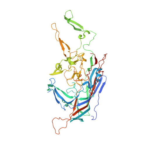
Structural characterization of the dual glycan binding adeno-associated virus serotype 6.
Ng, R., Govindasamy, L., Gurda, B.L., McKenna, R., Kozyreva, O.G., Samulski, R.J., Parent, K.N., Baker, T.S., Agbandje-McKenna, M.(2010) J Virol 84: 12945-12957
- PubMed: 20861247
- DOI: https://doi.org/10.1128/JVI.01235-10
- Primary Citation of Related Structures:
3OAH - PubMed Abstract:
The three-dimensional structure of adeno-associated virus (AAV) serotype 6 (AAV6) was determined using cryo-electron microscopy and image reconstruction and using X-ray crystallography to 9.7- and 3.0-Å resolution, respectively. The AAV6 capsid contains a highly conserved, eight-stranded (βB to βI) β-barrel core and large loop regions between the strands which form the capsid surface, as observed in other AAV structures. The loops show conformational variation compared to other AAVs, consistent with previous reports that amino acids in these loop regions are involved in differentiating AAV receptor binding, transduction efficiency, and antigenicity properties. Toward structure-function annotation of AAV6 with respect to its unique dual glycan receptor (heparan sulfate and sialic acid) utilization for cellular recognition, and its enhanced lung epithelial transduction compared to other AAVs, the capsid structure was compared to that of AAV1, which binds sialic acid and differs from AAV6 in only 6 out of 736 amino acids. Five of these residues are located at or close to the icosahedral 3-fold axis of the capsid, thereby identifying this region as imparting important functions, such as receptor attachment and transduction phenotype. Two of the five observed amino acids are located in the capsid interior, suggesting that differential AAV infection properties are also controlled by postentry intracellular events. Density ordered inside the capsid, under the 3-fold axis in a previously reported, conserved AAV DNA binding pocket, was modeled as a nucleotide and a base, further implicating this capsid region in AAV genome recognition and/or stabilization.
Organizational Affiliation:
Department of Biochemistry and Molecular Biology, University of Florida, Gainesville, FL 32610-0266, USA.