3O55

Crystal structure of human FAD-linked augmenter of liver regeneration (ALR)


Experimental Data Snapshot

  • Method: X-RAY DIFFRACTION
  • Resolution: 1.90 Å
  • R-Value Free: 0.267 
  • R-Value Work: 0.182 
  • R-Value Observed: 0.189 

wwPDB Validation   3D Report Full Report


Ligand Structure Quality Assessment 


This is version 1.3 of the entry. See complete history


Literature

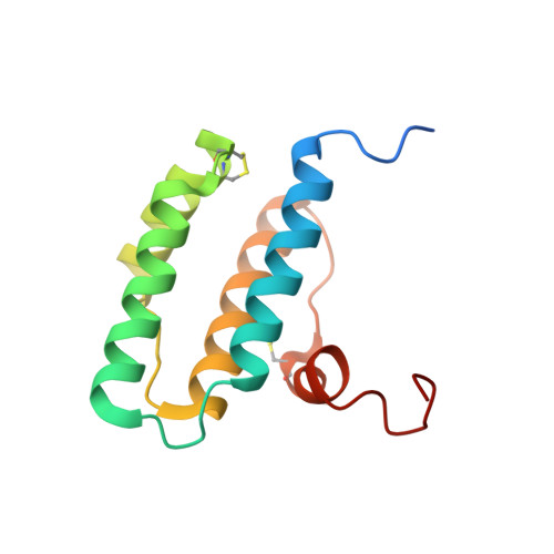
Molecular recognition and substrate mimicry drive the electron-transfer process between MIA40 and ALR.

Banci, L.Bertini, I.Calderone, V.Cefaro, C.Ciofi-Baffoni, S.Gallo, A.Kallergi, E.Lionaki, E.Pozidis, C.Tokatlidis, K.

(2011) Proc Natl Acad Sci U S A 108: 4811-4816

  • DOI: https://doi.org/10.1073/pnas.1014542108
  • Primary Citation of Related Structures:  
    3O55

  • PubMed Abstract: 

    Oxidative protein folding in the mitochondrial intermembrane space requires the transfer of a disulfide bond from MIA40 to the substrate. During this process MIA40 is reduced and regenerated to a functional state through the interaction with the flavin-dependent sulfhydryl oxidase ALR. Here we present the mechanistic basis of ALR-MIA40 interaction at atomic resolution by biochemical and structural analyses of the mitochondrial ALR isoform and its covalent mixed disulfide intermediate with MIA40. This ALR isoform contains a folded FAD-binding domain at the C-terminus and an unstructured, flexible N-terminal domain, weakly and transiently interacting one with the other. A specific region of the N-terminal domain guides the interaction with the MIA40 substrate binding cleft (mimicking the interaction of the substrate itself), without being involved in the import of ALR. The hydrophobicity-driven binding of this region ensures precise protein-protein recognition needed for an efficient electron transfer process.


  • Organizational Affiliation

    Magnetic Resonance Center (CERM), University of Florence, Via Luigi Sacconi 6, Sesto Fiorentino, 50019 Florence, Italy. banci@cerm.unifi.it


Macromolecules
Find similar proteins by:  (by identity cutoff)  |  3D Structure
Entity ID: 1
MoleculeChains Sequence LengthOrganismDetailsImage
Augmenter of liver regeneration125Homo sapiensMutation(s): 2 
Gene Names: ALR
UniProt & NIH Common Fund Data Resources
Find proteins for P55789 (Homo sapiens)
Explore P55789 
Go to UniProtKB:  P55789
PHAROS:  P55789
GTEx:  ENSG00000127554 
Entity Groups  
Sequence Clusters30% Identity50% Identity70% Identity90% Identity95% Identity100% Identity
UniProt GroupP55789
Sequence Annotations
Expand
  • Reference Sequence
Small Molecules
Ligands 1 Unique
IDChains Name / Formula / InChI Key2D Diagram3D Interactions
FAD
Query on FAD

Download Ideal Coordinates CCD File 
B [auth A]FLAVIN-ADENINE DINUCLEOTIDE
C27 H33 N9 O15 P2
VWWQXMAJTJZDQX-UYBVJOGSSA-N
Experimental Data & Validation

Experimental Data

  • Method: X-RAY DIFFRACTION
  • Resolution: 1.90 Å
  • R-Value Free: 0.267 
  • R-Value Work: 0.182 
  • R-Value Observed: 0.189 
  • Space Group: C 2 2 21
Unit Cell:
Length ( Å )Angle ( ˚ )
a = 50.849α = 90
b = 76.566β = 90
c = 62.31γ = 90
Software Package:
Software NamePurpose
CrysalisProdata collection
MOLREPphasing
REFMACrefinement
MOSFLMdata reduction
SCALAdata scaling

Structure Validation

View Full Validation Report


Ligand Structure Quality Assessment 


Entry History 

Deposition Data

Revision History  (Full details and data files)

  • Version 1.0: 2011-04-13
    Type: Initial release
  • Version 1.1: 2011-07-13
    Changes: Version format compliance
  • Version 1.2: 2017-11-08
    Changes: Refinement description
  • Version 1.3: 2023-09-06
    Changes: Data collection, Database references, Derived calculations, Refinement description