3NQS

Crystal Structure of Inducible Nitric Oxide Synthase with N-Nitrosated-pterin


Experimental Data Snapshot

  • Method: X-RAY DIFFRACTION
  • Resolution: 2.20 Å
  • R-Value Free: 0.229 
  • R-Value Work: 0.204 
  • R-Value Observed: 0.204 

wwPDB Validation   3D Report Full Report


Ligand Structure Quality Assessment 


This is version 1.3 of the entry. See complete history


Literature

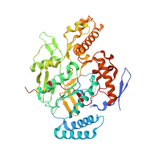
Nitric-oxide synthase forms N-NO-pterin and S-NO-cys: implications for activity, allostery, and regulation.

Rosenfeld, R.J.Bonaventura, J.Szymczyna, B.R.MacCoss, M.J.Arvai, A.S.Yates, J.R.Tainer, J.A.Getzoff, E.D.

(2010) J Biol Chem 285: 31581-31589

  • DOI: https://doi.org/10.1074/jbc.M109.072496
  • Primary Citation of Related Structures:  
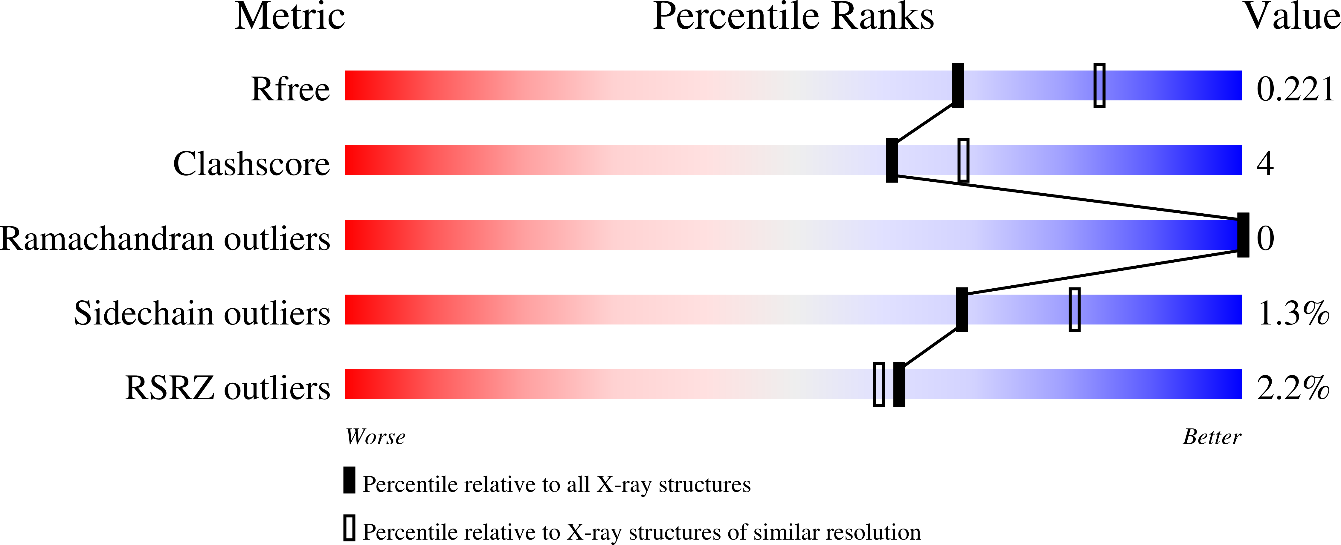
    3NQS

  • PubMed Abstract: 

    Inducible nitric-oxide synthase (iNOS) produces biologically stressful levels of nitric oxide (NO) as a potent mediator of cellular cytotoxicity or signaling. Yet, how this nitrosative stress affects iNOS function in vivo is poorly understood. Here we define two specific non-heme iNOS nitrosation sites discovered by combining UV-visible spectroscopy, chemiluminescence, mass spectrometry, and x-ray crystallography. We detected auto-S-nitrosylation during enzymatic turnover by using chemiluminescence. Selective S-nitrosylation of the ZnS(4) site, which bridges the dimer interface, promoted a dimer-destabilizing order-to-disorder transition. The nitrosated iNOS crystal structure revealed an unexpected N-NO modification on the pterin cofactor. Furthermore, the structurally defined N-NO moiety is solvent-exposed and available to transfer NO to a partner. We investigated glutathione (GSH) as a potential transnitrosation partner because the intracellular GSH concentration is high and NOS can form S-nitrosoglutathione. Our computational results predicted a GSH binding site adjacent to the N-NO-pterin. Moreover, we detected GSH binding to iNOS with saturation transfer difference NMR spectroscopy. Collectively, these observations resolve previous paradoxes regarding this uncommon pterin cofactor in NOS and suggest means for regulating iNOS activity via N-NO-pterin and S-NO-Cys modifications. The iNOS self-nitrosation characterized here appears appropriate to help control NO production in response to cellular conditions.


  • Organizational Affiliation

    Department of Molecular Biology, The Skaggs Institute for Chemical Biology, La Jolla, California 92037, USA.


Macromolecules
Find similar proteins by:  (by identity cutoff)  |  3D Structure
Entity ID: 1
MoleculeChains Sequence LengthOrganismDetailsImage
Nitric oxide synthase, inducible
A, B
433Mus musculusMutation(s): 0 
Gene Names: InoslNos2
EC: 1.14.13.39
UniProt & NIH Common Fund Data Resources
Find proteins for P29477 (Mus musculus)
Explore P29477 
Go to UniProtKB:  P29477
IMPC:  MGI:97361
Entity Groups  
Sequence Clusters30% Identity50% Identity70% Identity90% Identity95% Identity100% Identity
UniProt GroupP29477
Sequence Annotations
Expand
  • Reference Sequence
Small Molecules
Ligands 8 Unique
IDChains Name / Formula / InChI Key2D Diagram3D Interactions
HEM
Query on HEM

Download Ideal Coordinates CCD File 
C [auth A],
M [auth B]
PROTOPORPHYRIN IX CONTAINING FE
C34 H32 Fe N4 O4
KABFMIBPWCXCRK-RGGAHWMASA-L
BOG
Query on BOG

Download Ideal Coordinates CCD File 
R [auth B]octyl beta-D-glucopyranoside
C14 H28 O6
HEGSGKPQLMEBJL-RKQHYHRCSA-N
AT2
Query on AT2

Download Ideal Coordinates CCD File 
F [auth A],
P [auth B]
ETHYL 4-[(4-METHYLPYRIDIN-2-YL)AMINO]PIPERIDINE-1-CARBOXYLATE
C14 H21 N3 O2
LNRMJBWADUSJTA-UHFFFAOYSA-N
H4B
Query on H4B

Download Ideal Coordinates CCD File 
D [auth A],
N [auth B]
5,6,7,8-TETRAHYDROBIOPTERIN
C9 H15 N5 O3
FNKQXYHWGSIFBK-RPDRRWSUSA-N
SO4
Query on SO4

Download Ideal Coordinates CCD File 
J [auth A],
K [auth A],
L [auth A],
S [auth B],
T [auth B]
SULFATE ION
O4 S
QAOWNCQODCNURD-UHFFFAOYSA-L
GOL
Query on GOL

Download Ideal Coordinates CCD File 
G [auth A]GLYCEROL
C3 H8 O3
PEDCQBHIVMGVHV-UHFFFAOYSA-N
EDO
Query on EDO

Download Ideal Coordinates CCD File 
H [auth A],
I [auth A],
Q [auth B]
1,2-ETHANEDIOL
C2 H6 O2
LYCAIKOWRPUZTN-UHFFFAOYSA-N
NO
Query on NO

Download Ideal Coordinates CCD File 
E [auth A],
O [auth B]
NITRIC OXIDE
N O
ODUCDPQEXGNKDN-UHFFFAOYSA-N
Binding Affinity Annotations 
IDSourceBinding Affinity
AT2 BindingDB:  3NQS IC50: 350 (nM) from 1 assay(s)
Experimental Data & Validation

Experimental Data

  • Method: X-RAY DIFFRACTION
  • Resolution: 2.20 Å
  • R-Value Free: 0.229 
  • R-Value Work: 0.204 
  • R-Value Observed: 0.204 
  • Space Group: P 61 2 2
Unit Cell:
Length ( Å )Angle ( ˚ )
a = 214.114α = 90
b = 214.114β = 90
c = 116.542γ = 120
Software Package:
Software NamePurpose
ADSCdata collection
XFITdata reduction
CNSrefinement
DENZOdata reduction
HKL-2000data scaling

Structure Validation

View Full Validation Report


Ligand Structure Quality Assessment 


Entry History 

Deposition Data

Revision History  (Full details and data files)

  • Version 1.0: 2010-07-21
    Type: Initial release
  • Version 1.1: 2011-07-13
    Changes: Version format compliance
  • Version 1.2: 2020-07-29
    Type: Remediation
    Reason: Carbohydrate remediation
    Changes: Data collection, Derived calculations, Structure summary
  • Version 1.3: 2023-09-06
    Changes: Data collection, Database references, Refinement description, Structure summary