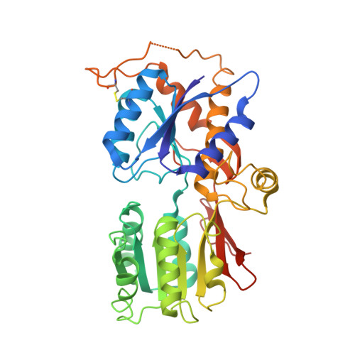
Subunit-selective N-terminal domain associations organize the formation of AMPA receptor heteromers
Rossmann, M., Sukumaran, M., Penn, A.C., Veprintsev, D.B., Babu, M.M., Greger, I.H.(2011) EMBO J 30: 959-971
- PubMed: 21317873
- DOI: https://doi.org/10.1038/emboj.2011.16
- Primary Citation of Related Structures:
3HSY, 3N6V, 3O2J - PubMed Abstract:
The assembly of AMPA-type glutamate receptors (AMPARs) into distinct ion channel tetramers ultimately governs the nature of information transfer at excitatory synapses. How cells regulate the formation of diverse homo- and heteromeric AMPARs is unknown. Using a sensitive biophysical approach, we show that the extracellular, membrane-distal AMPAR N-terminal domains (NTDs) orchestrate selective routes of heteromeric assembly via a surprisingly wide spectrum of subunit-specific association affinities. Heteromerization is dominant, occurs at the level of the dimer, and results in a preferential incorporation of the functionally critical GluA2 subunit. Using a combination of structure-guided mutagenesis and electrophysiology, we further map evolutionarily variable hotspots in the NTD dimer interface, which modulate heteromerization capacity. This 'flexibility' of the NTD not only explains why heteromers predominate but also how GluA2-lacking, Ca(2+)-permeable homomers could form, which are induced under specific physiological and pathological conditions. Our findings reveal that distinct NTD properties set the stage for the biogenesis of functionally diverse pools of homo- and heteromeric AMPAR tetramers.
Organizational Affiliation:
Neurobiology Division, MRC Laboratory of Molecular Biology, Cambridge, UK.


