Near-atomic resolution structures of urate oxidase complexed with its substrate and analogues: the protonation state of the ligand.
Gabison, L., Chiadmi, M., El Hajji, M., Castro, B., Colloc'h, N., Prange, T.(2010) Acta Crystallogr D Biol Crystallogr 66: 714-724
- PubMed: 20516624
- DOI: https://doi.org/10.1107/S090744491001142X
- Primary Citation of Related Structures:
3L8W, 3L9G, 3LBG, 3LD4 - PubMed Abstract:
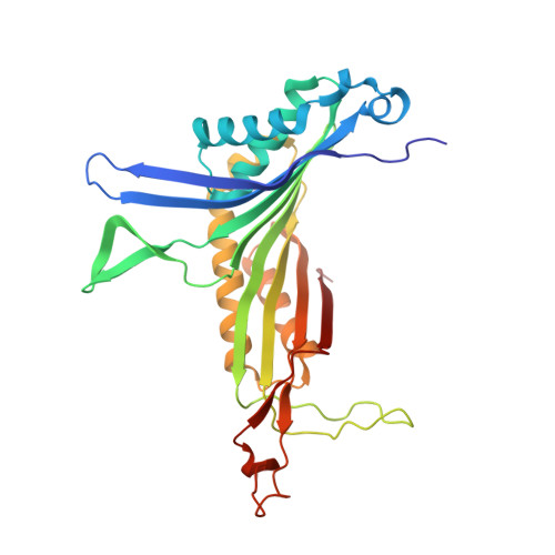
Urate oxidase (uricase; EC 1.7.3.3; UOX) from Aspergillus flavus catalyzes the oxidation of uric acid in the presence of molecular oxygen to 5-hydroxyisourate in the degradation cascade of purines; intriguingly, catalysis proceeds using neither a metal ion (Fe, Cu etc.) nor a redox cofactor. UOX is a tetrameric enzyme with four active sites located at the interface of two subunits; its structure was refined at atomic resolution (1 A) using new crystal data in the presence of xanthine and at near-atomic resolution (1.3-1.7 A) in complexes with the natural substrate (urate) and two inhibitors: 8-nitroxanthine and 8-thiouric acid. Three new features of the structural and mechanistic behaviour of the enzyme were addressed. Firstly, the high resolution of the UOX-xanthine structure allowed the solution of an old structural problem at a contact zone within the tetramer; secondly, the protonation state of the substrate was determined from both a halochromic inhibitor complex (UOX-8-nitroxanthine) and from the H-atom distribution in the active site, using the structures of the UOX-xanthine and the UOX-uric acid complexes; and thirdly, it was possible to extend the general base system, characterized by the conserved catalytic triad Thr-Lys-His, to a large water network that is able to buffer and shuttle protons back and forth between the substrate and the peroxo hole along the reaction pathway.
Organizational Affiliation:
Laboratoire de Cristallographie et RMN Biologiques, UMR 8015 CNRS, France.