3JWA

Crystal structure of L-methionine gamma-lyase from Citrobacter freundii with methionine phosphinate


Experimental Data Snapshot

  • Method: X-RAY DIFFRACTION
  • Resolution: 1.45 Å
  • R-Value Free: 0.182 
  • R-Value Work: 0.141 
  • R-Value Observed: 0.143 

wwPDB Validation   3D Report Full Report


Ligand Structure Quality Assessment 


This is version 1.5 of the entry. See complete history


Literature

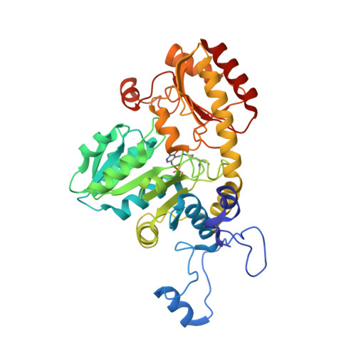
Three-dimensional structures of noncovalent complexes of Citrobacter freundii methionine gamma-lyase with substrates.

Revtovich, S.V.Morozova, E.A.Khurs, E.N.Zakomirdina, L.N.Nikulin, A.D.Demidkina, T.V.Khomutov, R.M.

(2011) Biochemistry (Mosc) 76: 564-570

  • DOI: https://doi.org/10.1134/S0006297911050063
  • Primary Citation of Related Structures:  
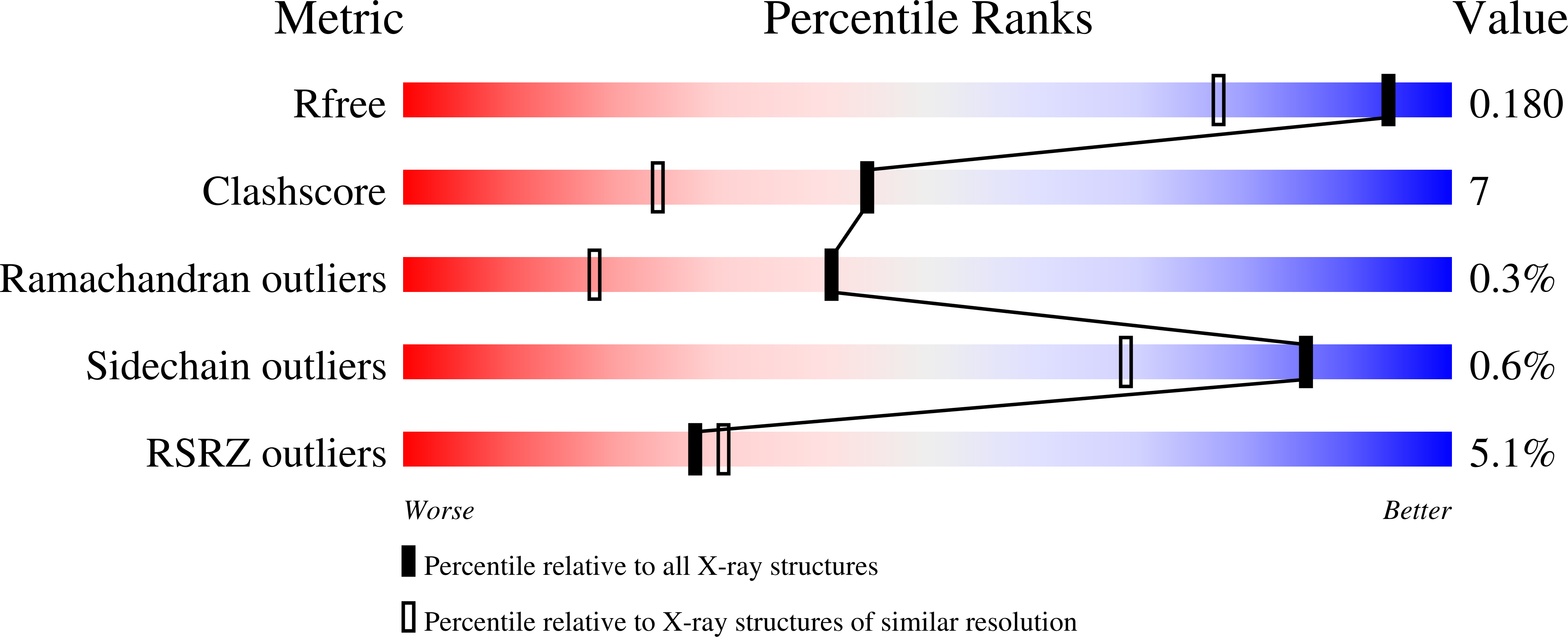
    3JW9, 3JWA, 3JWB

  • PubMed Abstract: 

    Crystal structures of Citrobacter freundii methionine γ-lyase complexes with the substrates of γ- (L-1-amino-3-methylthiopropylphosphinic acid) and β- (S-ethyl-L-cysteine) elimination reactions and the competitive inhibitor L-norleucine have been determined at 1.45, 1.8, and 1.63 Å resolution, respectively. All three amino acids occupy the active site of the enzyme but do not form a covalent bond with pyridoxal 5'-phosphate. Hydrophobic interactions between the active site residues and the side groups of the substrates and the inhibitor are supposed to cause noncovalent binding. Arg374 and Ser339 are involved in the binding of carboxyl groups of the substrates and the inhibitor. The hydroxyl of Tyr113 is a potential acceptor of a proton from the amino groups of the amino acids.


  • Organizational Affiliation

    Engelhardt Institute of Molecular Biology, Russian Academy of Sciences, Moscow. tvd@eimb.ru


Macromolecules
Find similar proteins by:  (by identity cutoff)  |  3D Structure
Entity ID: 1
MoleculeChains Sequence LengthOrganismDetailsImage
Methionine gamma-lyase398Citrobacter freundiiMutation(s): 0 
Gene Names: megL
EC: 4.4.1.11
UniProt
Find proteins for Q84AR1 (Citrobacter freundii)
Explore Q84AR1 
Go to UniProtKB:  Q84AR1
Entity Groups  
Sequence Clusters30% Identity50% Identity70% Identity90% Identity95% Identity100% Identity
UniProt GroupQ84AR1
Sequence Annotations
Expand
  • Reference Sequence
Experimental Data & Validation

Experimental Data

  • Method: X-RAY DIFFRACTION
  • Resolution: 1.45 Å
  • R-Value Free: 0.182 
  • R-Value Work: 0.141 
  • R-Value Observed: 0.143 
  • Space Group: I 2 2 2
Unit Cell:
Length ( Å )Angle ( ˚ )
a = 56.42α = 90
b = 123.01β = 90
c = 127.01γ = 90
Software Package:
Software NamePurpose
MAR345dtbdata collection
PHENIXmodel building
PHENIXrefinement
XDSdata reduction
XSCALEdata scaling
PHENIXphasing

Structure Validation

View Full Validation Report


Ligand Structure Quality Assessment 


Entry History 

Deposition Data

Revision History  (Full details and data files)

  • Version 1.0: 2010-09-08
    Type: Initial release
  • Version 1.1: 2011-07-13
    Changes: Version format compliance
  • Version 1.2: 2014-11-05
    Changes: Database references
  • Version 1.3: 2020-10-21
    Changes: Data collection, Derived calculations
  • Version 1.4: 2023-09-06
    Changes: Data collection, Database references, Refinement description
  • Version 1.5: 2023-11-22
    Changes: Data collection