3H5K

Crystal structure of the ribosome inactivating protein PDL1


Experimental Data Snapshot

  • Method: X-RAY DIFFRACTION
  • Resolution: 1.45 Å
  • R-Value Free: 0.242 
  • R-Value Work: 0.196 
  • R-Value Observed: 0.198 

wwPDB Validation   3D Report Full Report


Ligand Structure Quality Assessment 


This is version 1.2 of the entry. See complete history


Literature

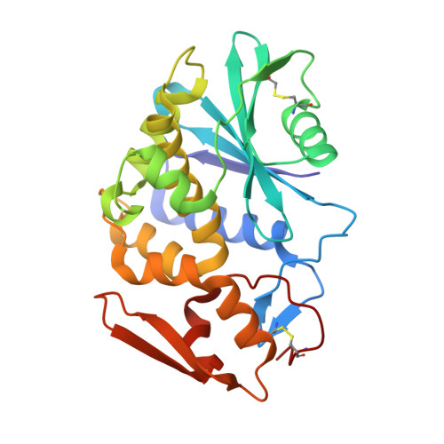
Crystal structure of PD-L1, a ribosome inactivating protein from Phytolacca dioica L. Leaves with the property to induce DNA cleavage

Ruggiero, A.Di Maro, A.Severino, V.Chambery, A.Berisio, R.

(2009) Biopolymers 91: 1135-1142

  • DOI: https://doi.org/10.1002/bip.21260
  • Primary Citation of Related Structures:  
    3H5K

  • PubMed Abstract: 

    The structure of the highly glycosylated type 1 ribosome inactivating protein PD-L1 was determined by X-ray crystallography. This protein belongs to a group of four PD-Ls (PD-L1-4) expressed in Phytolacca dioica leaves. Of these, PD-L1 and PD-L2 are endowed with the ability to cleave double strand DNA, a property which is not shared by the other two components of the family. Single crystals of native PD-L1, the most glycosylated, were obtained using seeding techniques and phase determination was achieved using molecular replacement. To investigate the role of glycosylation in the different functionality of these proteins, we performed DNA cleavage assays on the E. coli plasmid pBR322. These experiments revealed that DNA cleaving ability does not depend on the level of glycosylation of PD-L1, since there is no difference in the activities displayed by native PD-L1 and a recombinant non-glycosylated form. Besides, confirming that DNA cleavage by PD-L1 cannot be attributed to contaminations, these data unambiguously show that functional changes between PD-L1 and PD-L4 are solely to be attributed to their sequence differences. On the basis of the comparison of PD-L1 and PD-L4 crystal structures, we propose possible structural determinants responsible for their different functional behavior.


  • Organizational Affiliation

    Istituto di Biostrutture e Bioimmagini, Napoli I-80134, Italy.


Macromolecules
Find similar proteins by:  (by identity cutoff)  |  3D Structure
Entity ID: 1
MoleculeChains Sequence LengthOrganismDetailsImage
Ribosome-inactivating protein PD-L1/PD-L2
A, B
261Phytolacca dioicaMutation(s): 0 
EC: 3.2.2.22
UniProt
Find proteins for P84853 (Phytolacca dioica)
Explore P84853 
Go to UniProtKB:  P84853
Entity Groups  
Sequence Clusters30% Identity50% Identity70% Identity90% Identity95% Identity100% Identity
UniProt GroupP84853
Sequence Annotations
Expand
  • Reference Sequence
Experimental Data & Validation

Experimental Data

  • Method: X-RAY DIFFRACTION
  • Resolution: 1.45 Å
  • R-Value Free: 0.242 
  • R-Value Work: 0.196 
  • R-Value Observed: 0.198 
  • Space Group: C 1 2 1
Unit Cell:
Length ( Å )Angle ( ˚ )
a = 161.011α = 90
b = 34.735β = 127.99
c = 120.63γ = 90
Software Package:
Software NamePurpose
HKL-2000data collection
REMOmodel building
REFMACrefinement
HKL-2000data reduction
HKL-2000data scaling
REMOphasing

Structure Validation

View Full Validation Report


Ligand Structure Quality Assessment 


Entry History 

Deposition Data

Revision History  (Full details and data files)

  • Version 1.0: 2009-10-13
    Type: Initial release
  • Version 1.1: 2011-07-13
    Changes: Non-polymer description, Version format compliance
  • Version 1.2: 2020-07-29
    Type: Remediation
    Reason: Carbohydrate remediation
    Changes: Data collection, Derived calculations, Structure summary