3FK7

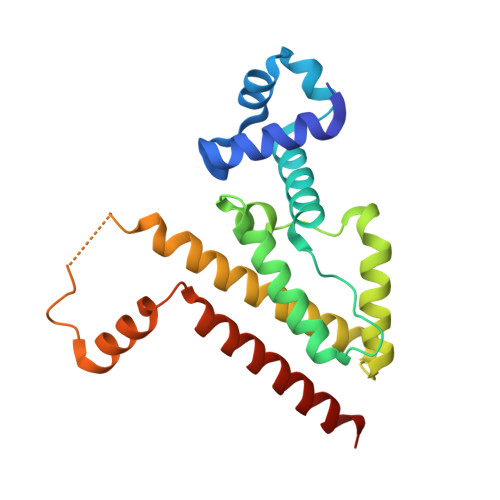
Crystal structure of TetR triple mutant (H64K, S135L, S138I) in complex with 4-ddma-atc


Experimental Data Snapshot

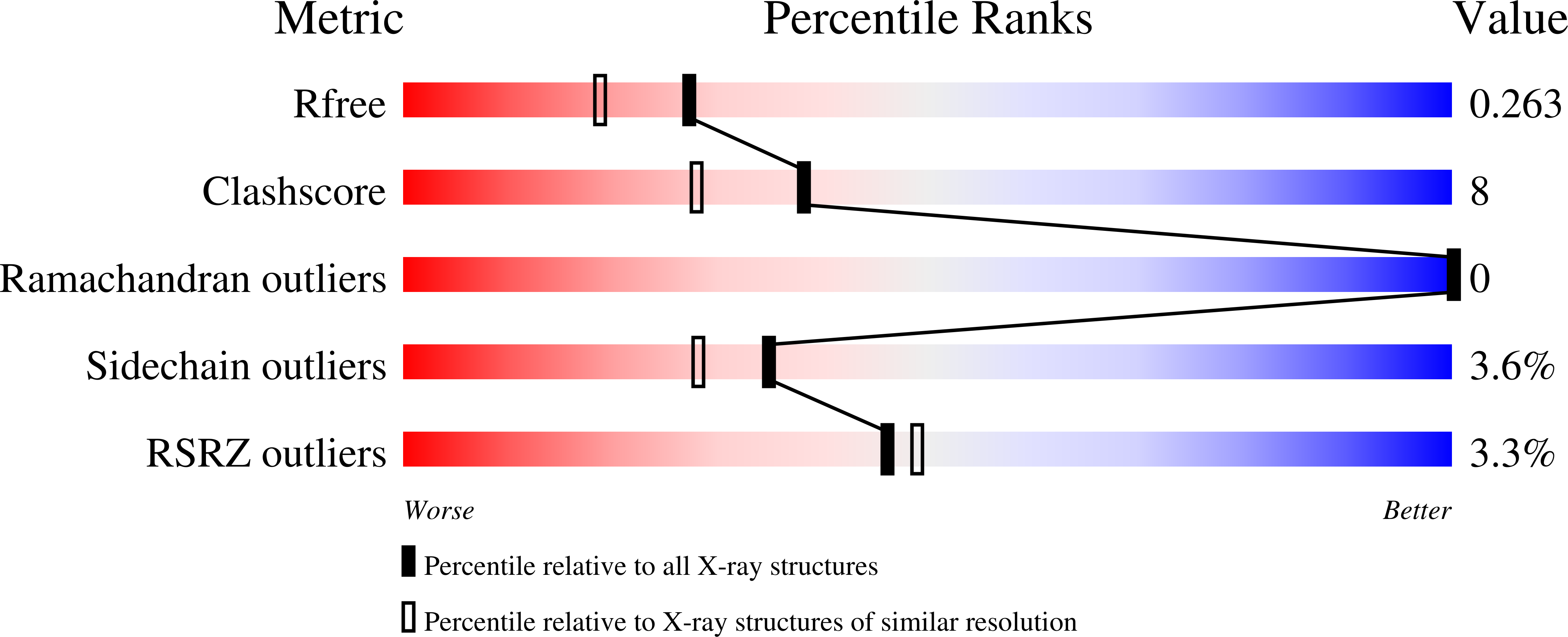
  • Method: X-RAY DIFFRACTION
  • Resolution: 2.06 Å
  • R-Value Free: 0.261 
  • R-Value Work: 0.203 
  • R-Value Observed: 0.208 

wwPDB Validation   3D Report Full Report


Ligand Structure Quality Assessment 


This is version 1.5 of the entry. See complete history


Literature

Structural origins for selectivity and specificity in an engineered bacterial repressor-inducer pair.

Klieber, M.A.Scholz, O.Lochner, S.Gmeiner, P.Hillen, W.Muller, Y.A.

(2009) FEBS J 276: 5610-5621

  • DOI: https://doi.org/10.1111/j.1742-4658.2009.07254.x
  • Primary Citation of Related Structures:  
    3FK6, 3FK7

  • PubMed Abstract: 

    The bacterial tetracycline transcription regulation system mediated by the tetracycline repressor (TetR) is widely used to study gene expression in prokaryotes and eukaryotes. To study multiple genes in parallel, a triple mutant TetR(K(64)L(135)I(138)) has been engineered that is selectively induced by the synthetic tetracycline derivative 4-de-dimethylamino-anhydrotetracycline (4-ddma-atc) and no longer by tetracycline, the inducer of wild-type TetR. In the present study, we report the crystal structure of TetR(K(64)L(135)I(138)) in the absence and in complex with 4-ddma-atc at resolutions of 2.1 A. Analysis of the structures in light of the available binding data and previously reported TetR complexes allows for a dissection of the origins of selectivity and specificity. In all crystal structures solved to date, the ligand-binding position, as well as the positioning of the residues lining the binding site, is extremely well conserved, irrespective of the chemical nature of the ligand. Selective recognition of 4-ddma-atc is achieved through fine-tuned hydrogen-bonding constraints introduced by the His64-->Lys substitution, as well as a combination of hydrophobic effect and the removal of unfavorable electrostatic interactions through the introduction of Leu135 and Ile138.


  • Organizational Affiliation

    Department of Biology, Lehrstuhl für Biotechnik, Friedrich-Alexander University, Erlangen-Nuremberg, Germany.


Macromolecules
Find similar proteins by:  (by identity cutoff)  |  3D Structure
Entity ID: 1
MoleculeChains Sequence LengthOrganismDetailsImage
Tetracycline repressor protein class B from transposon Tn10, Tetracycline repressor protein class D
A, B
208Escherichia coliMutation(s): 3 
Gene Names: tetR
UniProt
Find proteins for P0ACT4 (Escherichia coli)
Explore P0ACT4 
Go to UniProtKB:  P0ACT4
Find proteins for P04483 (Escherichia coli)
Explore P04483 
Go to UniProtKB:  P04483
Entity Groups  
Sequence Clusters30% Identity50% Identity70% Identity90% Identity95% Identity100% Identity
UniProt GroupsP0ACT4P04483
Sequence Annotations
Expand
  • Reference Sequence
Small Molecules
Binding Affinity Annotations 
IDSourceBinding Affinity
4DM Binding MOAD:  3FK7 Kd: 0.75 (nM) from 1 assay(s)
Experimental Data & Validation

Experimental Data

  • Method: X-RAY DIFFRACTION
  • Resolution: 2.06 Å
  • R-Value Free: 0.261 
  • R-Value Work: 0.203 
  • R-Value Observed: 0.208 
  • Space Group: C 1 2 1
Unit Cell:
Length ( Å )Angle ( ˚ )
a = 130.22α = 90
b = 59.38β = 97.85
c = 63.78γ = 90
Software Package:
Software NamePurpose
REFMACrefinement
PDB_EXTRACTdata extraction
MAR345dtbdata collection
XDSdata reduction
XSCALEdata scaling
AMoREphasing

Structure Validation

View Full Validation Report


Ligand Structure Quality Assessment 


Entry History 

Deposition Data

Revision History  (Full details and data files)

  • Version 1.0: 2009-11-03
    Type: Initial release
  • Version 1.1: 2011-07-13
    Changes: Version format compliance
  • Version 1.2: 2017-08-23
    Changes: Refinement description, Source and taxonomy
  • Version 1.3: 2017-11-01
    Changes: Refinement description
  • Version 1.4: 2021-11-10
    Changes: Database references, Derived calculations
  • Version 1.5: 2023-11-01
    Changes: Data collection, Refinement description