3EX8

Complex structure of bacillus subtilis RibG reduction mechanism in riboflavin biosynthesis


Experimental Data Snapshot

  • Method: X-RAY DIFFRACTION
  • Resolution: 2.56 Å
  • R-Value Free: 0.283 
  • R-Value Work: 0.229 
  • R-Value Observed: 0.229 

wwPDB Validation   3D Report Full Report


Ligand Structure Quality Assessment 


This is version 1.2 of the entry. See complete history


Literature

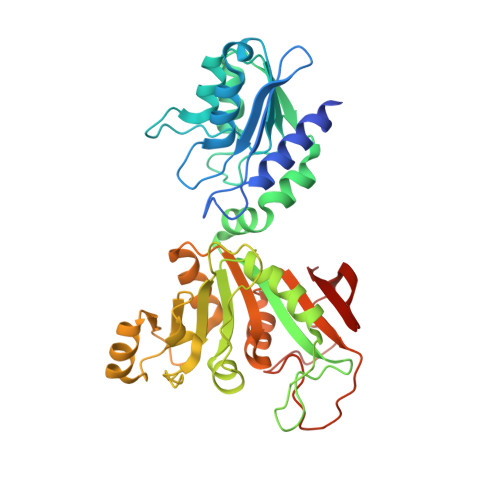
Complex structure of Bacillus subtilis RibG: the reduction mechanism during riboflavin biosynthesis.

Chen, S.C.Lin, Y.H.Yu, H.C.Liaw, S.H.

(2009) J Biol Chem 284: 1725-1731

  • DOI: https://doi.org/10.1074/jbc.M805820200
  • Primary Citation of Related Structures:  
    3EX8

  • PubMed Abstract: 

    Bacterial RibG is a potent target for antimicrobial agents, because it catalyzes consecutive deamination and reduction steps in the riboflavin biosynthesis. In the N-terminal deaminase domain of Bacillus subtilis RibG, structure-based mutational analyses demonstrated that Glu51 and Lys79 are essential for the deaminase activity. In the C-terminal reductase domain, the complex structure with the substrate at 2.56-A resolution unexpectedly showed a ribitylimino intermediate bound at the active site, and hence suggested that the ribosyl reduction occurs through a Schiff base pathway. Lys151 seems to have evolved to ensure specific recognition of the deaminase product rather than the substrate. Glu290, instead of the previously proposed Asp199, would seem to assist in the proton transfer in the reduction reaction. A detailed comparison reveals that the reductase and the pharmaceutically important enzyme, dihydrofolate reductase involved in the riboflavin and folate biosyntheses, share strong conservation of the core structure, cofactor binding, catalytic mechanism, even the substrate binding architecture.


  • Organizational Affiliation

    Structural Biology Program, National Yang-Ming University, Taipei 11221, Taiwan.


Macromolecules
Find similar proteins by:  (by identity cutoff)  |  3D Structure
Entity ID: 1
MoleculeChains Sequence LengthOrganismDetailsImage
Riboflavin biosynthesis protein ribD
A, B, C, D
373Bacillus subtilisMutation(s): 0 
Gene Names: BSU23280ribDribG
EC: 3.5.4.26 (PDB Primary Data), 1.1.1.193 (PDB Primary Data)
UniProt
Find proteins for P17618 (Bacillus subtilis (strain 168))
Explore P17618 
Go to UniProtKB:  P17618
Entity Groups  
Sequence Clusters30% Identity50% Identity70% Identity90% Identity95% Identity100% Identity
UniProt GroupP17618
Sequence Annotations
Expand
  • Reference Sequence
Experimental Data & Validation

Experimental Data

  • Method: X-RAY DIFFRACTION
  • Resolution: 2.56 Å
  • R-Value Free: 0.283 
  • R-Value Work: 0.229 
  • R-Value Observed: 0.229 
  • Space Group: P 21 21 21
Unit Cell:
Length ( Å )Angle ( ˚ )
a = 87.078α = 90
b = 108.367β = 90
c = 187.394γ = 90
Software Package:
Software NamePurpose
HKL-2000data collection
CNSrefinement
HKL-2000data reduction
HKL-2000data scaling
CNSphasing

Structure Validation

View Full Validation Report


Ligand Structure Quality Assessment 


Entry History 

Deposition Data

Revision History  (Full details and data files)

  • Version 1.0: 2008-11-04
    Type: Initial release
  • Version 1.1: 2011-07-13
    Changes: Version format compliance
  • Version 1.2: 2023-11-01
    Changes: Data collection, Database references, Derived calculations, Refinement description