3DHG

Crystal Structure of Toluene 4-Monoxygenase Hydroxylase


Experimental Data Snapshot

  • Method: X-RAY DIFFRACTION
  • Resolution: 1.85 Å
  • R-Value Free: 0.214 
  • R-Value Work: 0.164 
  • R-Value Observed: 0.167 

wwPDB Validation   3D Report Full Report


This is version 1.5 of the entry. See complete history


Literature

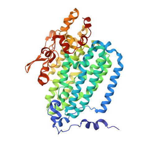
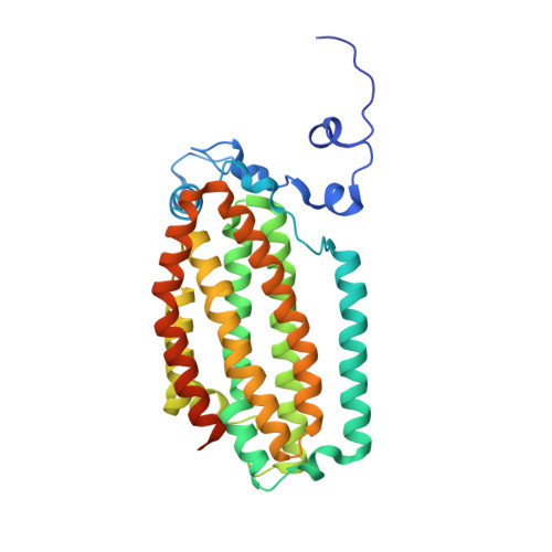
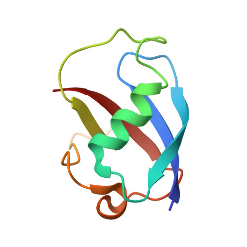
Structural consequences of effector protein complex formation in a diiron hydroxylase.

Bailey, L.J.McCoy, J.G.Phillips Jr., G.N.Fox, B.G.

(2008) Proc Natl Acad Sci U S A 105: 19194-19198

  • DOI: https://doi.org/10.1073/pnas.0807948105
  • Primary Citation of Related Structures:  
    3DHG, 3DHH, 3DHI

  • PubMed Abstract: 

    Carboxylate-bridged diiron hydroxylases are multicomponent enzyme complexes responsible for the catabolism of a wide range of hydrocarbons and as such have drawn attention for their mechanism of action and potential uses in bioremediation and enzymatic synthesis. These enzyme complexes use a small molecular weight effector protein to modulate the function of the hydroxylase. However, the origin of these functional changes is poorly understood. Here, we report the structures of the biologically relevant effector protein-hydroxylase complex of toluene 4-monooxygenase in 2 redox states. The structures reveal a number of coordinated changes that occur up to 25 A from the active site and poise the diiron center for catalysis. The results provide a structural basis for the changes observed in a number of the measurable properties associated with effector protein binding. This description provides insight into the functional role of effector protein binding in all carboxylate-bridged diiron hydroxylases.


  • Organizational Affiliation

    Department of Biochemistry, University of Wisconsin, 433 Babcock Drive, Madison, WI 53706-1544, USA.


Macromolecules
Find similar proteins by:  (by identity cutoff)  |  3D Structure
Entity ID: 1
MoleculeChains Sequence LengthOrganismDetailsImage
toluene 4-monooxygenase hydroxylase alpha subunit
A, D
500Pseudomonas mendocinaMutation(s): 0 
Gene Names: tmoA
UniProt
Find proteins for Q00456 (Pseudomonas mendocina)
Explore Q00456 
Go to UniProtKB:  Q00456
Entity Groups  
Sequence Clusters30% Identity50% Identity70% Identity90% Identity95% Identity100% Identity
UniProt GroupQ00456
Sequence Annotations
Expand
  • Reference Sequence
Find similar proteins by:  (by identity cutoff)  |  3D Structure
Entity ID: 2
MoleculeChains Sequence LengthOrganismDetailsImage
toluene 4-monooxygenase hydroxylase beta subunit
B, E
327Pseudomonas mendocinaMutation(s): 0 
Gene Names: tmoE
UniProt
Find proteins for Q00460 (Pseudomonas mendocina)
Explore Q00460 
Go to UniProtKB:  Q00460
Entity Groups  
Sequence Clusters30% Identity50% Identity70% Identity90% Identity95% Identity100% Identity
UniProt GroupQ00460
Sequence Annotations
Expand
  • Reference Sequence
Find similar proteins by:  (by identity cutoff)  |  3D Structure
Entity ID: 3
MoleculeChains Sequence LengthOrganismDetailsImage
toluene 4-monooxygenase hydroxylase gamma subunit
C, F
84Pseudomonas mendocinaMutation(s): 0 
Gene Names: tmoB
EC: 1.14.13
UniProt
Find proteins for Q00457 (Pseudomonas mendocina)
Explore Q00457 
Go to UniProtKB:  Q00457
Entity Groups  
Sequence Clusters30% Identity50% Identity70% Identity90% Identity95% Identity100% Identity
UniProt GroupQ00457
Sequence Annotations
Expand
  • Reference Sequence
Small Molecules
Ligands 3 Unique
IDChains Name / Formula / InChI Key2D Diagram3D Interactions
FE
Query on FE

Download Ideal Coordinates CCD File 
G [auth A],
H [auth A],
O [auth D],
P [auth D]
FE (III) ION
Fe
VTLYFUHAOXGGBS-UHFFFAOYSA-N
AZI
Query on AZI

Download Ideal Coordinates CCD File 
Q [auth D]AZIDE ION
N3
IVRMZWNICZWHMI-UHFFFAOYSA-N
CA
Query on CA

Download Ideal Coordinates CCD File 
I [auth B]
J [auth B]
K [auth B]
L [auth B]
M [auth B]
I [auth B],
J [auth B],
K [auth B],
L [auth B],
M [auth B],
N [auth C],
R [auth E],
S [auth E],
T [auth F]
CALCIUM ION
Ca
BHPQYMZQTOCNFJ-UHFFFAOYSA-N
Experimental Data & Validation

Experimental Data

  • Method: X-RAY DIFFRACTION
  • Resolution: 1.85 Å
  • R-Value Free: 0.214 
  • R-Value Work: 0.164 
  • R-Value Observed: 0.167 
  • Space Group: P 1 21 1
Unit Cell:
Length ( Å )Angle ( ˚ )
a = 56.822α = 90
b = 181.481β = 107.62
c = 89.917γ = 90
Software Package:
Software NamePurpose
DENZOdata reduction
SCALEPACKdata scaling
MOLREPphasing
REFMACrefinement
PDB_EXTRACTdata extraction

Structure Validation

View Full Validation Report


Entry History 

Deposition Data

Revision History  (Full details and data files)

  • Version 1.0: 2008-12-30
    Type: Initial release
  • Version 1.1: 2011-07-13
    Changes: Version format compliance
  • Version 1.2: 2013-01-30
    Changes: Structure summary
  • Version 1.3: 2016-12-14
    Changes: Structure summary
  • Version 1.4: 2017-10-25
    Changes: Refinement description
  • Version 1.5: 2024-02-21
    Changes: Data collection, Database references, Derived calculations