3DEZ

Crystal structure of Orotate phosphoribosyltransferase from Streptococcus mutans


Experimental Data Snapshot

  • Method: X-RAY DIFFRACTION
  • Resolution: 2.40 Å
  • R-Value Free: 0.249 
  • R-Value Work: 0.218 
  • R-Value Observed: 0.218 

wwPDB Validation   3D Report Full Report


This is version 1.3 of the entry. See complete history


Literature

Structure of orotate phosphoribosyltransferase from the caries pathogen Streptococcus mutans

Liu, C.P.Xu, R.Gao, Z.Q.Xu, J.H.Hou, H.F.Li, L.Q.She, Z.Li, L.F.Su, X.D.Liu, P.Dong, Y.H.

(2010) Acta Crystallogr Sect F Struct Biol Cryst Commun 66: 498-502

  • DOI: https://doi.org/10.1107/S1744309110009243
  • Primary Citation of Related Structures:  
    3DEZ

  • PubMed Abstract: 

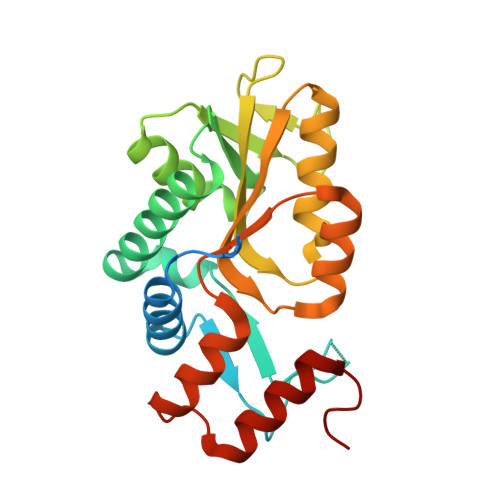
    Orotate phosphoribosyltransferase (OPRTase) catalyzes the OMP-forming step in de novo pyrimidine-nucleotide biosynthesis. Here, the crystal structure of OPRTase from the caries pathogen Streptococcus mutans is reported at 2.4 A resolution. S. mutans OPRTase forms a symmetric dimer and each monomer binds two sulfates at the active sites. The structural symmetry of the sulfate-binding sites and the missing loops in this structure are consistent with a symmetric catalysis mechanism.


  • Organizational Affiliation

    Beijing Synchrotron Radiation Facility, Institute of High Energy Physics, Chinese Academy of Sciences, Beijing 100049, People's Republic of China.


Macromolecules
Find similar proteins by:  (by identity cutoff)  |  3D Structure
Entity ID: 1
MoleculeChains Sequence LengthOrganismDetailsImage
Orotate phosphoribosyltransferase
A, B
243Streptococcus mutansMutation(s): 1 
Gene Names: pyrE
EC: 2.4.2.10
UniProt
Find proteins for Q8DTV2 (Streptococcus mutans serotype c (strain ATCC 700610 / UA159))
Explore Q8DTV2 
Go to UniProtKB:  Q8DTV2
Entity Groups  
Sequence Clusters30% Identity50% Identity70% Identity90% Identity95% Identity100% Identity
UniProt GroupQ8DTV2
Sequence Annotations
Expand
  • Reference Sequence
Experimental Data & Validation

Experimental Data

  • Method: X-RAY DIFFRACTION
  • Resolution: 2.40 Å
  • R-Value Free: 0.249 
  • R-Value Work: 0.218 
  • R-Value Observed: 0.218 
  • Space Group: P 41 21 2
Unit Cell:
Length ( Å )Angle ( ˚ )
a = 73.798α = 90
b = 73.798β = 90
c = 184.779γ = 90
Software Package:
Software NamePurpose
HKL-2000data collection
PHASESphasing
CNSrefinement
HKL-2000data reduction
HKL-2000data scaling

Structure Validation

View Full Validation Report


Entry History 

Deposition Data

Revision History  (Full details and data files)

  • Version 1.0: 2009-06-16
    Type: Initial release
  • Version 1.1: 2011-07-13
    Changes: Version format compliance
  • Version 1.2: 2021-11-10
    Changes: Database references, Derived calculations
  • Version 1.3: 2023-11-01
    Changes: Data collection, Refinement description