


Structure of the E. coli transcriptional repressor ascG.
Singer, A.U., Kagan, O., Evdokimova, E., Osipiuk, J., Edwards, A.M., Joachimiak, A., Savchenko, A.To be published.
Experimental Data Snapshot
Entity ID: 1 | |||||
|---|---|---|---|---|---|
| Molecule | Chains | Sequence Length | Organism | Details | Image |
| HTH-type transcriptional regulator ascG | 296 | Escherichia coli K-12 | Mutation(s): 0 Gene Names: ascG, b2714, JW5434 | 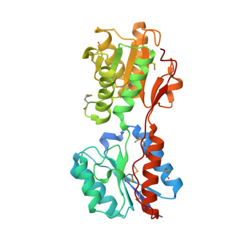 | |
UniProt | |||||
Find proteins for P24242 (Escherichia coli (strain K12)) Explore P24242 Go to UniProtKB: P24242 | |||||
Entity Groups | |||||
| Sequence Clusters | 30% Identity50% Identity70% Identity90% Identity95% Identity100% Identity | ||||
| UniProt Group | P24242 | ||||
Sequence AnnotationsExpand | |||||
| |||||
| Ligands 3 Unique | |||||
|---|---|---|---|---|---|
| ID | Chains | Name / Formula / InChI Key | 2D Diagram | 3D Interactions | |
| FRU Query on FRU | C [auth A] | beta-D-fructofuranose C6 H12 O6 RFSUNEUAIZKAJO-ARQDHWQXSA-N |  | ||
| SO4 Query on SO4 | D [auth A], F [auth B] | SULFATE ION O4 S QAOWNCQODCNURD-UHFFFAOYSA-L |  | ||
| NA Query on NA | E [auth B] | SODIUM ION Na FKNQFGJONOIPTF-UHFFFAOYSA-N |  | ||
| Modified Residues 1 Unique | |||||
|---|---|---|---|---|---|
| ID | Chains | Type | Formula | 2D Diagram | Parent |
| MSE Query on MSE | A, B | L-PEPTIDE LINKING | C5 H11 N O2 Se |  | MET |
| Length ( Å ) | Angle ( ˚ ) |
|---|---|
| a = 62.229 | α = 90 |
| b = 65.446 | β = 90 |
| c = 130.706 | γ = 90 |
| Software Name | Purpose |
|---|---|
| REFMAC | refinement |
| ADSC | data collection |
| HKL-2000 | data reduction |
| HKL-2000 | data scaling |
| HKL-3000 | phasing |
RCSB PDB (citation) is hosted by
RCSB PDB is a member of the

