Molecular basis for defect in Alix-binding by alternatively spliced isoform of ALG-2 (ALG-2DeltaGF122) and structural roles of F122 in target recognition
Inuzuka, T., Suzuki, H., Kawasaki, M., Shibata, H., Wakatsuki, S., Maki, M.(2010) BMC Struct Biol 10: 25-25
- PubMed: 20691033
- DOI: https://doi.org/10.1186/1472-6807-10-25
- Primary Citation of Related Structures:
3AAJ, 3AAK - PubMed Abstract:
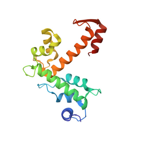
ALG-2 (a gene product of PDCD6) belongs to the penta-EF-hand (PEF) protein family and Ca2+-dependently interacts with various intracellular proteins including mammalian Alix, an adaptor protein in the ESCRT system. Our previous X-ray crystal structural analyses revealed that binding of Ca2+ to EF3 enables the side chain of R125 to move enough to make a primary hydrophobic pocket (Pocket 1) accessible to a short fragment of Alix. The side chain of F122, facing a secondary hydrophobic pocket (Pocket 2), interacts with the Alix peptide. An alternatively spliced shorter isoform, designated ALG-2DeltaGF122, lacks Gly121Phe122 and does not bind Alix, but the structural basis of the incompetence has remained to be elucidated. We solved the X-ray crystal structure of the PEF domain of ALG-2DeltaGF122 in the Ca2+-bound form and compared it with that of ALG-2. Deletion of the two residues shortened alpha-helix 5 (alpha5) and changed the configuration of the R125 side chain so that it partially blocked Pocket 1. A wall created by the main chain of 121-GFG-123 and facing the two pockets was destroyed. Surprisingly, however, substitution of F122 with Ala or Gly, but not with Trp, increased the Alix-binding capacity in binding assays. The F122 substitutions exhibited different effects on binding of ALG-2 to other known interacting proteins, including TSG101 (Tumor susceptibility gene 101) and annexin A11. The X-ray crystal structure of the F122A mutant revealed that removal of the bulky F122 side chain not only created an additional open space in Pocket 2 but also abolished inter-helix interactions with W95 and V98 (present in alpha4) and that alpha5 inclined away from alpha4 to expand Pocket 2, suggesting acquirement of more appropriate positioning of the interacting residues to accept Alix. We found that the inability of the two-residue shorter ALG-2 isoform to bind Alix is not due to the absence of bulky side chain of F122 but due to deformation of a main-chain wall facing pockets 1 and 2. Moreover, a residue at the position of F122 contributes to target specificity and a smaller side chain is preferable for Alix binding but not favored to bind annexin A11.
Organizational Affiliation:
Department of Applied Molecular Biosciences, Graduate School of Bioagricultural Sciences, Nagoya University, Nagoya 464-8601, Japan.