2I5R

Structure of small Toprim domain-containing protein from B. stearothermophilus in complex with Mg2+


Experimental Data Snapshot

  • Method: X-RAY DIFFRACTION
  • Resolution: 1.65 Å
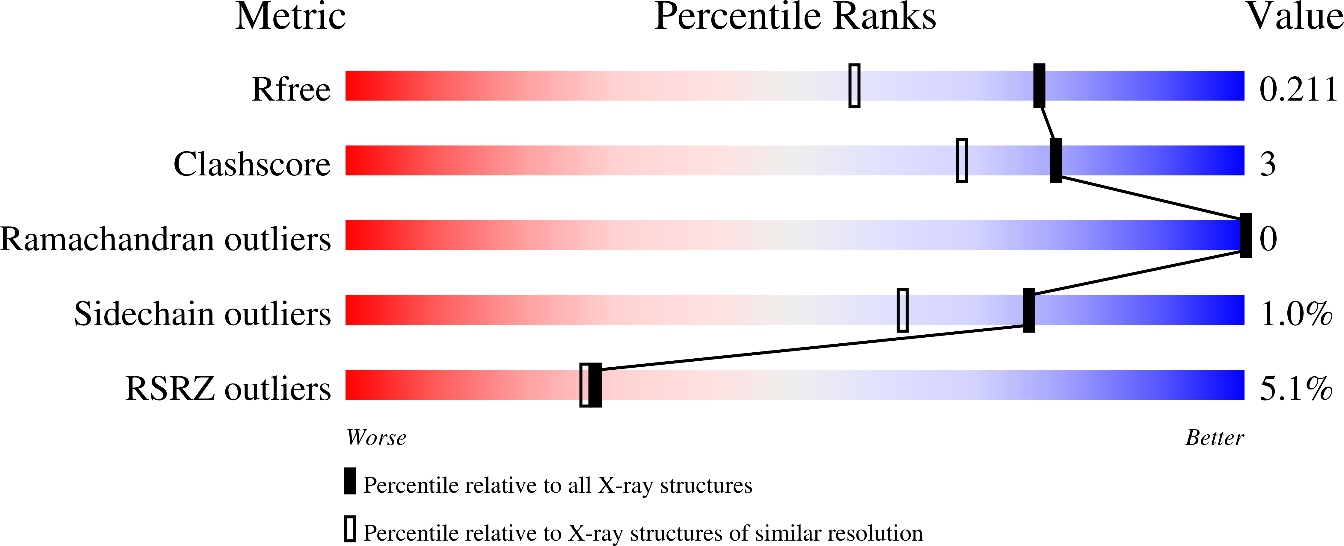
  • R-Value Free: 0.208 
  • R-Value Work: 0.171 
  • R-Value Observed: 0.173 

wwPDB Validation   3D Report Full Report


This is version 1.4 of the entry. See complete history


Literature

Crystal structure and putative function of small Toprim domain-containing protein from Bacillus stearothermophilus.

Rezacova, P.Borek, D.Moy, S.F.Joachimiak, A.Otwinowski, Z.

(2007) Proteins 70: 311-319

  • DOI: https://doi.org/10.1002/prot.21511
  • Primary Citation of Related Structures:  
    2FCJ, 2I5R

  • PubMed Abstract: 

    The crystal structure of the Midwest Center for Structural Genomics target APC35832, a 14.7-kDa cytosolic protein from Bacillus stearothermophilus, has been determined at 1.3 A resolution by the single anomalous diffraction method from a mercury soaked crystal. The APC35832 protein is a representative of large group of bacterial and archeal proteins entirely consisting of the Toprim (topoisomerase-primase) domain. This domain is found in the catalytic centers of many enzymes catalyzing phosphodiester bond formation or cleavage, but the function of small Toprim domain proteins remains unknown. Consistent with the sequence analysis, the APC35832 structure shows a conserved Toprim fold, with a central 4-stranded parallel beta-sheet surrounded by four alpha-helixes. Comparison of the APC35832 structure with its closest structural homolog, the catalytic core of bacteriophage T7 primase, revealed structural conservation of a metal binding site and isothermal titration calorimetry indicates that APC35832 binds Mg2+ with a sub-millimolar dissociation constant (K(d)). The APC35832-Mg2+ complex structure was determined at 1.65 A and reveals the role of conserved acidic residues in Mg2+ ion coordination. The structural similarities to other Toprim domain containing proteins and potential function and substrates of APC35832 are discussed in this article.


  • Organizational Affiliation

    Department of Biochemistry, UT Southwestern Medical Center, Dallas, Texas 75390-8816, USA.


Macromolecules
Find similar proteins by:  (by identity cutoff)  |  3D Structure
Entity ID: 1
MoleculeChains Sequence LengthOrganismDetailsImage
Toprim domain-containing protein
A, B, C
122Geobacillus stearothermophilusMutation(s): 0 
UniProt
Find proteins for Q5KVJ9 (Geobacillus kaustophilus (strain HTA426))
Explore Q5KVJ9 
Go to UniProtKB:  Q5KVJ9
Entity Groups  
Sequence Clusters30% Identity50% Identity70% Identity90% Identity95% Identity100% Identity
UniProt GroupQ5KVJ9
Sequence Annotations
Expand
  • Reference Sequence
Small Molecules
Ligands 4 Unique
IDChains Name / Formula / InChI Key2D Diagram3D Interactions
MES
Query on MES

Download Ideal Coordinates CCD File 
L [auth C]2-(N-MORPHOLINO)-ETHANESULFONIC ACID
C6 H13 N O4 S
SXGZJKUKBWWHRA-UHFFFAOYSA-N
SO4
Query on SO4

Download Ideal Coordinates CCD File 
J [auth C],
K [auth C]
SULFATE ION
O4 S
QAOWNCQODCNURD-UHFFFAOYSA-L
GOL
Query on GOL

Download Ideal Coordinates CCD File 
E [auth A]
F [auth A]
H [auth B]
I [auth B]
M [auth C]
E [auth A],
F [auth A],
H [auth B],
I [auth B],
M [auth C],
N [auth C]
GLYCEROL
C3 H8 O3
PEDCQBHIVMGVHV-UHFFFAOYSA-N
MG
Query on MG

Download Ideal Coordinates CCD File 
D [auth A],
G [auth B]
MAGNESIUM ION
Mg
JLVVSXFLKOJNIY-UHFFFAOYSA-N
Experimental Data & Validation

Experimental Data

  • Method: X-RAY DIFFRACTION
  • Resolution: 1.65 Å
  • R-Value Free: 0.208 
  • R-Value Work: 0.171 
  • R-Value Observed: 0.173 
  • Space Group: P 21 21 21
Unit Cell:
Length ( Å )Angle ( ˚ )
a = 36.667α = 90
b = 73.031β = 90
c = 114.922γ = 90
Software Package:
Software NamePurpose
REFMACrefinement
HKL-2000data collection
HKL-2000data reduction
HKL-2000data scaling

Structure Validation

View Full Validation Report


Entry History 

Deposition Data

Revision History  (Full details and data files)

  • Version 1.0: 2007-07-03
    Type: Initial release
  • Version 1.1: 2007-10-11
    Changes: Version format compliance
  • Version 1.2: 2011-07-13
    Changes: Advisory, Version format compliance
  • Version 1.3: 2017-10-18
    Changes: Refinement description
  • Version 1.4: 2024-02-21
    Changes: Data collection, Database references, Derived calculations, Refinement description