2ZN8

Crystal structure of Zn2+-bound form of ALG-2


Experimental Data Snapshot

  • Method: X-RAY DIFFRACTION
  • Resolution: 2.70 Å
  • R-Value Free: 0.260 
  • R-Value Work: 0.211 
  • R-Value Observed: 0.213 

wwPDB Validation   3D Report Full Report


This is version 1.2 of the entry. See complete history


Literature

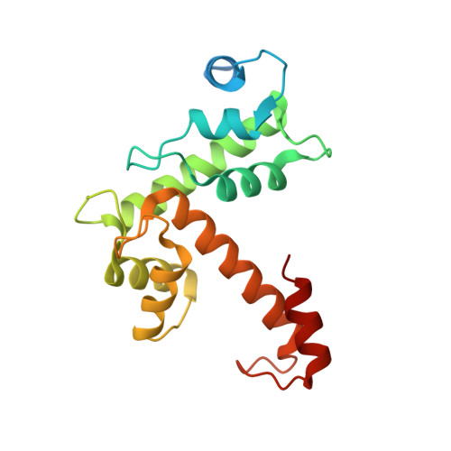
Structural Basis for Ca(2+)-Dependent Formation of ALG-2/Alix Peptide Complex: Ca(2+)/EF3-Driven Arginine Switch Mechanism

Suzuki, H.Kawasaki, M.Inuzuka, T.Okumura, M.Kakiuchi, T.Shibata, H.Wakatsuki, S.Maki, M.

(2008) Structure 16: 1562-1573

  • DOI: https://doi.org/10.1016/j.str.2008.07.012
  • Primary Citation of Related Structures:  
    2ZN8, 2ZN9, 2ZND, 2ZNE

  • PubMed Abstract: 

    ALG-2 belongs to the penta-EF-hand (PEF) protein family and interacts with various intracellular proteins, such as Alix and TSG101, that are involved in endosomal sorting and HIV budding. Through X-ray crystallography, we solved the structures of Ca(2+)-free and -bound forms of N-terminally truncated human ALG-2 (des3-20ALG-2), Zn(2+)-bound form of full-length ALG-2, and the structure of the complex between des3-23ALG-2 and the peptide corresponding to Alix799-814 in Zn(2+)-bound form. Binding of Ca(2+) to EF3 enables the side chain of Arg125, present in the loop connecting EF3 and EF4, to move enough to make a primary hydrophobic pocket accessible to the critical PPYP motif, which partially overlaps with the GPP motif for the binding of Cep55 (centrosome protein 55 kDa). Based on these results, together with the results of in vitro binding assay with mutant ALG-2 and Alix proteins, we propose a Ca(2+)/EF3-driven arginine switch mechanism for ALG-2 binding to Alix.


  • Organizational Affiliation

    Department of Applied Molecular Biosciences, Graduate School of Bioagricultural Sciences, Nagoya University, Nagoya 464-8601, Japan.


Macromolecules
Find similar proteins by:  (by identity cutoff)  |  3D Structure
Entity ID: 1
MoleculeChains Sequence LengthOrganismDetailsImage
Programmed cell death protein 6190Homo sapiensMutation(s): 0 
Gene Names: PDCD6ALG2
UniProt & NIH Common Fund Data Resources
Find proteins for O75340 (Homo sapiens)
Explore O75340 
Go to UniProtKB:  O75340
PHAROS:  O75340
GTEx:  ENSG00000249915 
Entity Groups  
Sequence Clusters30% Identity50% Identity70% Identity90% Identity95% Identity100% Identity
UniProt GroupO75340
Sequence Annotations
Expand
  • Reference Sequence
Experimental Data & Validation

Experimental Data

  • Method: X-RAY DIFFRACTION
  • Resolution: 2.70 Å
  • R-Value Free: 0.260 
  • R-Value Work: 0.211 
  • R-Value Observed: 0.213 
  • Space Group: C 2 2 21
Unit Cell:
Length ( Å )Angle ( ˚ )
a = 72.01α = 90
b = 169.738β = 90
c = 46.343γ = 90
Software Package:
Software NamePurpose
REFMACrefinement
HKL-2000data reduction
HKL-2000data scaling
MOLREPphasing

Structure Validation

View Full Validation Report


Entry History 

Deposition Data

Revision History  (Full details and data files)

  • Version 1.0: 2008-09-09
    Type: Initial release
  • Version 1.1: 2011-07-13
    Changes: Version format compliance
  • Version 1.2: 2023-11-01
    Changes: Data collection, Database references, Derived calculations, Refinement description