2Z1C

Crystal structure of HypC from Thermococcus kodakaraensis KOD1


Experimental Data Snapshot

  • Method: X-RAY DIFFRACTION
  • Resolution: 1.80 Å
  • R-Value Free: 0.246 
  • R-Value Work: 0.222 
  • R-Value Observed: 0.223 

wwPDB Validation   3D Report Full Report


This is version 1.3 of the entry. See complete history


Literature

Crystal Structures of [NiFe] Hydrogenase Maturation Proteins HypC, HypD, and HypE: Insights into Cyanation Reaction by Thiol Redox Signaling

Watanabe, S.Matsumi, R.Arai, T.Atomi, H.Imanaka, T.Miki, K.

(2007) Mol Cell 27: 29-40

  • DOI: https://doi.org/10.1016/j.molcel.2007.05.039
  • Primary Citation of Related Structures:  
    2Z1C, 2Z1D, 2Z1E, 2Z1F

  • PubMed Abstract: 

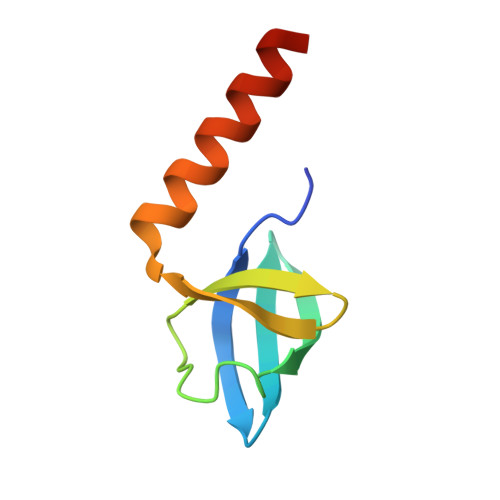
    [NiFe] hydrogenase maturation proteins HypC, HypD, and HypE catalyze the insertion and cyanation of the iron center of [NiFe] hydrogenases by an unknown mechanism. We have determined the crystal structures of HypC, HypD, and HypE from Thermococcus kodakaraensis KOD1 at 1.8 A, 2.07 A, and 1.55 A resolution, respectively. The structure of HypD reveals its probable iron binding and active sites for cyanation. An extended conformation of each conserved motif of HypC and HypE allows the essential cysteine residues of both proteins to interact with the active site of HypD. Furthermore, the C-terminal tail of HypE is shown to exist in an ATP-dependent dynamic equilibrium between outward and inward conformations. Unexpectedly, the [4Fe-4S] cluster environment of HypD is quite similar to that of ferredoxin:thioredoxin reductase (FTR), indicating the existence of a redox cascade similar to the FTR system. These results suggest a cyanation reaction mechanism via unique thiol redox signaling in the HypCDE complex.


  • Organizational Affiliation

    Department of Chemistry, Graduate School of Science, Kyoto University, Sakyo-ku, Kyoto 606-8502, Japan.


Macromolecules
Find similar proteins by:  (by identity cutoff)  |  3D Structure
Entity ID: 1
MoleculeChains Sequence LengthOrganismDetailsImage
Hydrogenase expression/formation protein HypC
A, B, C
75Thermococcus kodakarensis KOD1Mutation(s): 0 
Gene Names: hypC
UniProt
Find proteins for Q5JII0 (Thermococcus kodakarensis (strain ATCC BAA-918 / JCM 12380 / KOD1))
Explore Q5JII0 
Go to UniProtKB:  Q5JII0
Entity Groups  
Sequence Clusters30% Identity50% Identity70% Identity90% Identity95% Identity100% Identity
UniProt GroupQ5JII0
Sequence Annotations
Expand
  • Reference Sequence
Small Molecules
Experimental Data & Validation

Experimental Data

  • Method: X-RAY DIFFRACTION
  • Resolution: 1.80 Å
  • R-Value Free: 0.246 
  • R-Value Work: 0.222 
  • R-Value Observed: 0.223 
  • Space Group: C 1 2 1
Unit Cell:
Length ( Å )Angle ( ˚ )
a = 78.237α = 90
b = 59.13β = 109.01
c = 53.973γ = 90
Software Package:
Software NamePurpose
REFMACrefinement
HKL-2000data collection
HKL-2000data reduction
HKL-2000data scaling
SOLVEphasing

Structure Validation

View Full Validation Report


Entry History 

Deposition Data

Revision History  (Full details and data files)

  • Version 1.0: 2007-07-17
    Type: Initial release
  • Version 1.1: 2008-04-30
    Changes: Version format compliance
  • Version 1.2: 2011-07-13
    Changes: Advisory, Version format compliance
  • Version 1.3: 2024-03-13
    Changes: Data collection, Database references, Derived calculations