2YFO

GALACTOSIDASE DOMAIN OF ALPHA-GALACTOSIDASE-SUCROSE KINASE, AGASK, in complex with galactose


Experimental Data Snapshot

  • Method: X-RAY DIFFRACTION
  • Resolution: 1.35 Å
  • R-Value Free: 0.142 
  • R-Value Work: 0.124 
  • R-Value Observed: 0.125 

wwPDB Validation   3D Report Full Report


Ligand Structure Quality Assessment 


This is version 1.3 of the entry. See complete history


Literature

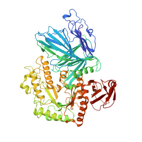
Agask, a Bifunctional Enzyme from the Human Microbiome Coupling Galactosidase and Kinase Activities

Bruel, L.Sulzenbacher, G.Tison-Cervera, M.Pujol, A.Nicoletti, C.Perrier, J.Galinier, A.Ropartz, D.Fons, M.Pompeo, F.Giardina, T.

(2011) J Biol Chem 286: 40814

  • DOI: https://doi.org/10.1074/jbc.M111.286039
  • Primary Citation of Related Structures:  
    2YFN, 2YFO

  • PubMed Abstract: 

    α-Galactosides are non-digestible carbohydrates widely distributed in plants. They are a potential source of energy in our daily food, and their assimilation by microbiota may play a role in obesity. In the intestinal tract, they are degraded by microbial glycosidases, which are often modular enzymes with catalytic domains linked to carbohydrate-binding modules. Here we introduce a bifunctional enzyme from the human intestinal bacterium Ruminococcus gnavus E1, α-galactosidase/sucrose kinase (AgaSK). Sequence analysis showed that AgaSK is composed of two domains: one closely related to α-galactosidases from glycoside hydrolase family GH36 and the other containing a nucleotide-binding motif. Its biochemical characterization showed that AgaSK is able to hydrolyze melibiose and raffinose to galactose and either glucose or sucrose, respectively, and to specifically phosphorylate sucrose on the C6 position of glucose in the presence of ATP. The production of sucrose-6-P directly from raffinose points toward a glycolytic pathway in bacteria, not described so far. The crystal structures of the galactosidase domain in the apo form and in complex with the product shed light onto the reaction and substrate recognition mechanisms and highlight an oligomeric state necessary for efficient substrate binding and suggesting a cross-talk between the galactose and kinase domains.


  • Organizational Affiliation

    Faculté des Sciences et Techniques Saint-Jérôme, Université Paul Cézanne, ISM2/BiosCiences UMR CNRS 6263, service 342, 13397 Marseille Cedex 20, France.


Macromolecules
Find similar proteins by:  (by identity cutoff)  |  3D Structure
Entity ID: 1
MoleculeChains Sequence LengthOrganismDetailsImage
ALPHA-GALACTOSIDASE-SUCROSE KINASE AGASK720Mediterraneibacter gnavus E1Mutation(s): 0 
EC: 3.2.1.22
UniProt
Find proteins for G4T4R7 (Ruminococcus gnavus)
Explore G4T4R7 
Go to UniProtKB:  G4T4R7
Entity Groups  
Sequence Clusters30% Identity50% Identity70% Identity90% Identity95% Identity100% Identity
UniProt GroupG4T4R7
Sequence Annotations
Expand
  • Reference Sequence
Small Molecules
Ligands 6 Unique
IDChains Name / Formula / InChI Key2D Diagram3D Interactions
GAL
Query on GAL

Download Ideal Coordinates CCD File 
JA [auth A]beta-D-galactopyranose
C6 H12 O6
WQZGKKKJIJFFOK-FPRJBGLDSA-N
GLA
Query on GLA

Download Ideal Coordinates CCD File 
IA [auth A]alpha-D-galactopyranose
C6 H12 O6
WQZGKKKJIJFFOK-PHYPRBDBSA-N
PGE
Query on PGE

Download Ideal Coordinates CCD File 
C [auth A],
D [auth A],
E [auth A],
F [auth A]
TRIETHYLENE GLYCOL
C6 H14 O4
ZIBGPFATKBEMQZ-UHFFFAOYSA-N
PO4
Query on PO4

Download Ideal Coordinates CCD File 
G [auth A]PHOSPHATE ION
O4 P
NBIIXXVUZAFLBC-UHFFFAOYSA-K
GOL
Query on GOL

Download Ideal Coordinates CCD File 
B [auth A]GLYCEROL
C3 H8 O3
PEDCQBHIVMGVHV-UHFFFAOYSA-N
NA
Query on NA

Download Ideal Coordinates CCD File 
AA [auth A]
BA [auth A]
CA [auth A]
DA [auth A]
EA [auth A]
AA [auth A],
BA [auth A],
CA [auth A],
DA [auth A],
EA [auth A],
FA [auth A],
GA [auth A],
H [auth A],
HA [auth A],
I [auth A],
J [auth A],
K [auth A],
L [auth A],
M [auth A],
N [auth A],
O [auth A],
P [auth A],
Q [auth A],
R [auth A],
S [auth A],
T [auth A],
U [auth A],
V [auth A],
W [auth A],
X [auth A],
Y [auth A],
Z [auth A]
SODIUM ION
Na
FKNQFGJONOIPTF-UHFFFAOYSA-N
Experimental Data & Validation

Experimental Data

  • Method: X-RAY DIFFRACTION
  • Resolution: 1.35 Å
  • R-Value Free: 0.142 
  • R-Value Work: 0.124 
  • R-Value Observed: 0.125 
  • Space Group: I 2 2 2
Unit Cell:
Length ( Å )Angle ( ˚ )
a = 105.016α = 90
b = 111.603β = 90
c = 155.29γ = 90
Software Package:
Software NamePurpose
REFMACrefinement
XDSdata reduction
SCALAdata scaling
REFMACphasing

Structure Validation

View Full Validation Report


Ligand Structure Quality Assessment 


Entry History 

Deposition Data

Revision History  (Full details and data files)

  • Version 1.0: 2011-09-28
    Type: Initial release
  • Version 1.1: 2011-11-23
    Changes: Database references, Derived calculations, Other
  • Version 1.2: 2011-12-07
    Changes: Database references
  • Version 1.3: 2020-07-29
    Type: Remediation
    Reason: Carbohydrate remediation
    Changes: Data collection, Derived calculations, Other, Structure summary