2Y9U

Structural basis of p63a SAM domain mutants involved in AEC syndrome


Experimental Data Snapshot

  • Method: X-RAY DIFFRACTION
  • Resolution: 1.60 Å
  • R-Value Free: 0.206 
  • R-Value Work: 0.185 
  • R-Value Observed: 0.186 

wwPDB Validation   3D Report Full Report


This is version 1.1 of the entry. See complete history


Literature

Structural Basis of P63Alpha Sam Domain Mutants Involved in Aec Syndrome.

Sathyamurthy, A.Freund, S.M.V.Johnson, C.M.Allen, M.D.Bycroft, M.

(2011) FEBS J 278: 2680

  • DOI: https://doi.org/10.1111/j.1742-4658.2011.08194.x
  • Primary Citation of Related Structures:  
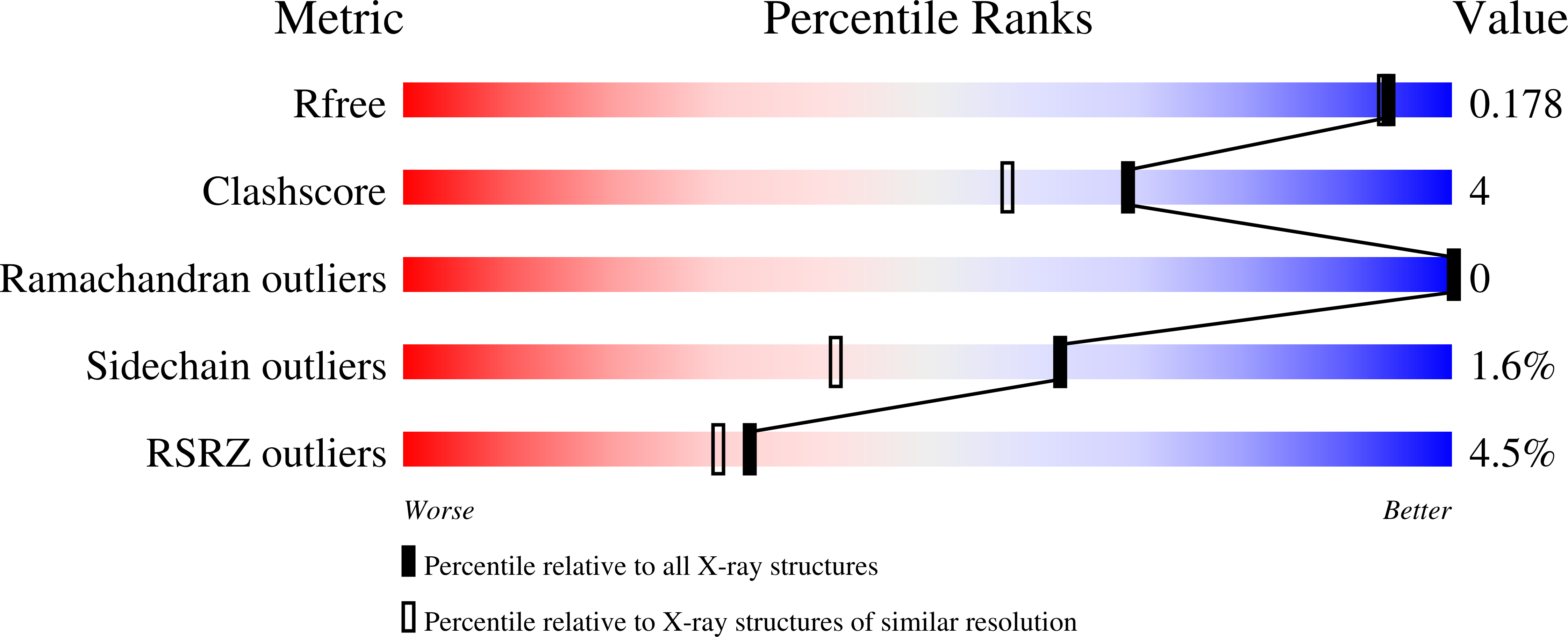
    2Y9T, 2Y9U

  • PubMed Abstract: 

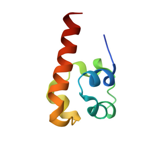
    p63 is a member of the p53 tumour suppressor family that includes p73. The p63 gene encodes a protein comprising an N-terminal transactivation domain, a DNA binding domain and an oligomerization domain, but varies in the organization of the C-terminus as a result of complex alternative splicing. p63α contains a C-terminal sterile α motif (SAM) domain that is thought to function as a protein-protein interaction domain. Several missense and heterozygous frame shift mutations, encoded within exon 13 and 14 of the p63 gene, have been identified in the p63α SAM domain in patients suffering from ankyloblepharon-ectodermal dysplasia-clefting syndrome. Here we report the solution and high resolution crystal structures of the p63α SAM domain and investigate the effect of several mutations (L553F/V, C562G/W, G569V, Q575L and I576T) on the stability of the domain. The possible effects of other mutations are also discussed.


  • Organizational Affiliation

    MRC Centre for Protein Engineering, Cambridge, UK.


Macromolecules
Find similar proteins by:  (by identity cutoff)  |  3D Structure
Entity ID: 1
MoleculeChains Sequence LengthOrganismDetailsImage
TUMOR PROTEIN 6369Homo sapiensMutation(s): 0 
UniProt & NIH Common Fund Data Resources
Find proteins for Q9H3D4 (Homo sapiens)
Explore Q9H3D4 
Go to UniProtKB:  Q9H3D4
PHAROS:  Q9H3D4
GTEx:  ENSG00000073282 
Entity Groups  
Sequence Clusters30% Identity50% Identity70% Identity90% Identity95% Identity100% Identity
UniProt GroupQ9H3D4
Sequence Annotations
Expand
  • Reference Sequence
Small Molecules
Ligands 1 Unique
IDChains Name / Formula / InChI Key2D Diagram3D Interactions
SO4
Query on SO4

Download Ideal Coordinates CCD File 
B [auth A]SULFATE ION
O4 S
QAOWNCQODCNURD-UHFFFAOYSA-L
Experimental Data & Validation

Experimental Data

  • Method: X-RAY DIFFRACTION
  • Resolution: 1.60 Å
  • R-Value Free: 0.206 
  • R-Value Work: 0.185 
  • R-Value Observed: 0.186 
  • Space Group: P 21 21 21
Unit Cell:
Length ( Å )Angle ( ˚ )
a = 34.624α = 90
b = 38.309β = 90
c = 44.516γ = 90
Software Package:
Software NamePurpose
PHENIXrefinement
MOSFLMdata reduction
SCALAdata scaling
PHASERphasing

Structure Validation

View Full Validation Report


Entry History 

Deposition Data

Revision History  (Full details and data files)

  • Version 1.0: 2011-08-03
    Type: Initial release
  • Version 1.1: 2018-01-24
    Changes: Source and taxonomy