2XNT

Acetylcholine binding protein (AChBP) as template for hierarchical in silico screening procedures to identify structurally novel ligands for the nicotinic receptors


Experimental Data Snapshot

  • Method: X-RAY DIFFRACTION
  • Resolution: 3.21 Å
  • R-Value Free: 0.232 
  • R-Value Work: 0.216 
  • R-Value Observed: 0.217 

wwPDB Validation   3D Report Full Report


Ligand Structure Quality Assessment 


This is version 1.3 of the entry. See complete history


Literature

Acetylcholine Binding Protein (Achbp) as Template for Hierarchical in Silico Screening Procedures to Identify Structurally Novel Ligands for the Nicotinic Receptors.

Akdemir, A.Rucktooa, P.Jongejan, A.Elk, R.V.Bertrand, S.Sixma, T.K.Bertrand, D.Smit, A.B.Leurs, R.De Graaf, C.De Esch, I.J.

(2011) Bioorg Med Chem 19: 6107

  • DOI: https://doi.org/10.1016/j.bmc.2011.08.028
  • Primary Citation of Related Structures:  
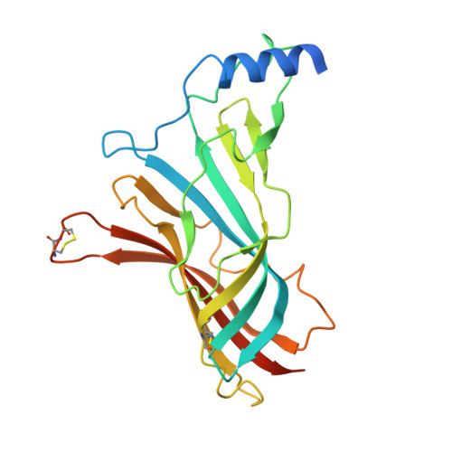
    2XNT, 2XNU, 2XNV

  • PubMed Abstract: 

    Hierarchical in silico screening protocols against the agonist bound acetylcholine binding protein (AChBP) crystal structure were efficient in identifying novel chemotypes for AChBP and the human α7 receptor. Two hit structures were cocrystallized with AChBP revealing intermolecular cation-π interactions with loop C but lacking intermolecular hydrogen bonding. The compounds act as competitive α7 receptor antagonists and as non-competitive α4β2 receptor inhibitors. These results underline the usability of AChBP in structure-based in silico screening strategies in finding novel scaffolds for the α7 receptor, but also illustrates some limitations of using AChBP as bait to find competitive α4β2 receptor ligands and α7 receptor agonists.


  • Organizational Affiliation

    Leiden/Amsterdam Center for Drug Research, Division of Medicinal Chemistry, Faculty of Sciences, VU University Amsterdam, The Netherlands.


Macromolecules
Find similar proteins by:  (by identity cutoff)  |  3D Structure
Entity ID: 1
MoleculeChains Sequence LengthOrganismDetailsImage
SOLUBLE ACETYLCHOLINE RECEPTOR
A, B, C, D, E
A, B, C, D, E, F, G, H, I, J
236Aplysia californicaMutation(s): 0 
UniProt
Find proteins for Q8WSF8 (Aplysia californica)
Explore Q8WSF8 
Go to UniProtKB:  Q8WSF8
Entity Groups  
Sequence Clusters30% Identity50% Identity70% Identity90% Identity95% Identity100% Identity
UniProt GroupQ8WSF8
Sequence Annotations
Expand
  • Reference Sequence
Small Molecules
Ligands 2 Unique
IDChains Name / Formula / InChI Key2D Diagram3D Interactions
VU2
Query on VU2

Download Ideal Coordinates CCD File 
AA [auth I]
CA [auth J]
K [auth A]
M [auth B]
O [auth C]
AA [auth I],
CA [auth J],
K [auth A],
M [auth B],
O [auth C],
Q [auth D],
S [auth E],
U [auth F],
W [auth G],
Y [auth H]
(2S)-2-[(4-CHLOROBENZYL)OXY]-2-PHENYLETHANAMINE
C15 H16 Cl N O
KFVPLCBTGAXYHI-OAHLLOKOSA-N
BR
Query on BR

Download Ideal Coordinates CCD File 
BA [auth I]
DA [auth J]
L [auth A]
N [auth B]
P [auth C]
BA [auth I],
DA [auth J],
L [auth A],
N [auth B],
P [auth C],
R [auth D],
T [auth E],
V [auth F],
X [auth G],
Z [auth H]
BROMIDE ION
Br
CPELXLSAUQHCOX-UHFFFAOYSA-M
Experimental Data & Validation

Experimental Data

  • Method: X-RAY DIFFRACTION
  • Resolution: 3.21 Å
  • R-Value Free: 0.232 
  • R-Value Work: 0.216 
  • R-Value Observed: 0.217 
  • Space Group: P 21 21 21
Unit Cell:
Length ( Å )Angle ( ˚ )
a = 129.111α = 90
b = 144.867β = 90
c = 145.106γ = 90
Software Package:
Software NamePurpose
BUSTERrefinement
XDSdata reduction
SCALAdata scaling
AMoREphasing

Structure Validation

View Full Validation Report


Ligand Structure Quality Assessment 


Entry History 

Deposition Data

Revision History  (Full details and data files)

  • Version 1.0: 2011-08-24
    Type: Initial release
  • Version 1.1: 2011-10-12
    Changes: Database references
  • Version 1.2: 2019-04-24
    Changes: Data collection, Other, Source and taxonomy
  • Version 1.3: 2023-12-20
    Changes: Data collection, Database references, Derived calculations, Other, Refinement description