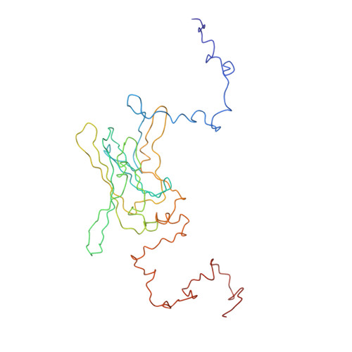
2XKY

Single particle analysis of Kir2.1NC_4 in negative stain


Experimental Data Snapshot

  • Method: ELECTRON MICROSCOPY
  • Resolution: 17.2 Å
  • Aggregation State: PARTICLE 
  • Reconstruction Method: SINGLE PARTICLE 

  • Method: SOLUTION SCATTERING

wwPDB Validation   3D Report Full Report


This is version 1.4 of the entry. See complete history


Literature

Self-Directed Assembly and Clustering of the Cytoplasmic Domains of Inwardly Rectifying Kir2.1 Potassium Channels on Association with Psd-95

Fomina, S.Howard, T.D.Sleator, O.K.Golovanova, M.O'Ryan, L.Leyland, M.L.Grossmann, J.G.Collins, R.F.Prince, S.M.

(2011) Biochim Biophys Acta 1808: 2374

  • DOI: https://doi.org/10.1016/j.bbamem.2011.06.021
  • Primary Citation of Related Structures:  
    2XKX, 2XKY

  • PubMed Abstract: 

    The interaction of the extra-membranous domain of tetrameric inwardly rectifying Kir2.1 ion channels (Kir2.1NC(4)) with the membrane associated guanylate kinase protein PSD-95 has been studied using Transmission Electron Microscopy in negative stain. Three types of complexes were observed in electron micrographs corresponding to a 1:1 complex, a large self-enclosed tetrad complex and extended chains of linked channel domains. Using models derived from small angle X-ray scattering experiments in which high resolution structures from X-ray crystallographic and Nuclear Magnetic Resonance studies are positioned, the envelopes from single particle analysis can be resolved as a Kir2.1NC(4):PSD-95 complex and a tetrad of this unit (Kir2.1NC(4):PSD-95)(4). The tetrad complex shows the close association of the Kir2.1 cytoplasmic domains and the influence of PSD-95 mediated self-assembly on the clustering of these channels.


  • Organizational Affiliation

    University of Manchester, Manchester Interdisciplinary Biocentre, Manchester M1 7DN, UK.


Macromolecules
Find similar proteins by:  (by identity cutoff)  |  3D Structure
Entity ID: 1
MoleculeChains Sequence LengthOrganismDetailsImage
INWARD RECTIFIER POTASSIUM CHANNEL 2A [auth I],
B [auth J],
C [auth K],
D [auth L]
309Mus musculusMutation(s): 0 
UniProt
Find proteins for P35561 (Mus musculus)
Explore P35561 
Go to UniProtKB:  P35561
Entity Groups  
Sequence Clusters30% Identity50% Identity70% Identity90% Identity95% Identity100% Identity
UniProt GroupP35561
Sequence Annotations
Expand
  • Reference Sequence
Experimental Data & Validation

Experimental Data

  • Method: ELECTRON MICROSCOPY
  • Resolution: 17.2 Å
  • Aggregation State: PARTICLE 
  • Reconstruction Method: SINGLE PARTICLE 
  • Method: SOLUTION SCATTERING
EM Software:
TaskSoftware PackageVersion
RECONSTRUCTIONEMAN

Structure Validation

View Full Validation Report


Entry History 

Deposition Data

Revision History  (Full details and data files)

  • Version 1.0: 2011-07-20
    Type: Initial release
  • Version 1.1: 2011-08-10
    Changes: Database references
  • Version 1.2: 2017-04-19
    Changes: Other
  • Version 1.3: 2017-08-30
    Changes: Data collection
  • Version 1.4: 2019-11-13
    Changes: Data collection, Other