2WBQ

Crystal structure of VioC in complex with (2S,3S)-hydroxyarginine


Experimental Data Snapshot

  • Method: X-RAY DIFFRACTION
  • Resolution: 1.10 Å
  • R-Value Free: 0.178 
  • R-Value Work: 0.143 
  • R-Value Observed: 0.144 

wwPDB Validation   3D Report Full Report


Ligand Structure Quality Assessment 


This is version 2.1 of the entry. See complete history


Literature

Structural Basis for the Erythro-Stereospecificity of the L-Arginine Oxygenase Vioc in Viomycin Biosynthesis.

Helmetag, V.Samel, S.A.Thomas, M.G.Marahiel, M.A.Essen, L.-O.

(2009) FEBS J 276: 3669

  • DOI: https://doi.org/10.1111/j.1742-4658.2009.07085.x
  • Primary Citation of Related Structures:  
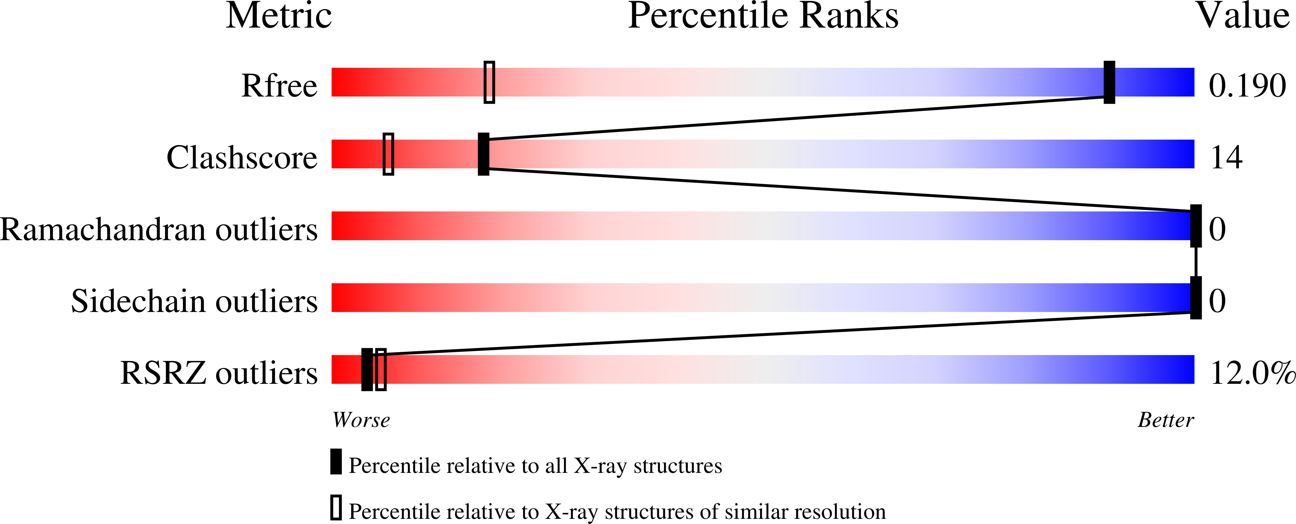
    2WBO, 2WBP, 2WBQ

  • PubMed Abstract: 

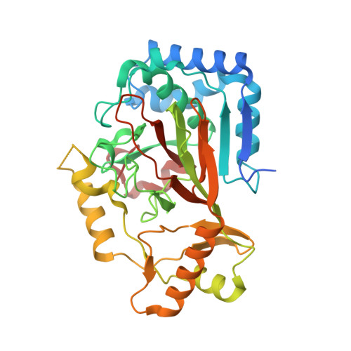
    The nonheme iron oxygenase VioC from Streptomyces vinaceus catalyzes Fe(II)-dependent and alpha-ketoglutarate-dependent Cbeta-hydroxylation of L-arginine during the biosynthesis of the tuberactinomycin antibiotic viomycin. Crystal structures of VioC were determined in complexes with the cofactor Fe(II), the substrate L-arginine, the product (2S,3S)-hydroxyarginine and the coproduct succinate at 1.1-1.3 A resolution. The overall structure reveals a beta-helix core fold with two additional helical subdomains that are common to nonheme iron oxygenases of the clavaminic acid synthase-like superfamily. In contrast to other clavaminic acid synthase-like oxygenases, which catalyze the formation of threo diastereomers, VioC produces the erythro diastereomer of Cbeta-hydroxylated L-arginine. This unexpected stereospecificity is caused by conformational control of the bound substrate, which enforces a gauche(-) conformer for chi(1) instead of the trans conformers observed for the asparagine oxygenase AsnO and other members of the clavaminic acid synthase-like superfamily. Additionally, the substrate specificity of VioC was investigated. The side chain of the L-arginine substrate projects outwards from the active site by undergoing interactions mainly with the C-terminal helical subdomain. Accordingly, VioC exerts broadened substrate specificity by accepting the analogs L-homoarginine and L-canavanine for Cbeta-hydroxylation.


  • Organizational Affiliation

    Department of Chemistry, Philipps-University Marburg, Germany.


Macromolecules
Find similar proteins by:  (by identity cutoff)  |  3D Structure
Entity ID: 1
MoleculeChains Sequence LengthOrganismDetailsImage
L-ARGININE BETA-HYDROXYLASE358Streptomyces vinaceusMutation(s): 0 
UniProt
Find proteins for Q6WZB0 (Streptomyces vinaceus)
Explore Q6WZB0 
Go to UniProtKB:  Q6WZB0
Entity Groups  
Sequence Clusters30% Identity50% Identity70% Identity90% Identity95% Identity100% Identity
UniProt GroupQ6WZB0
Sequence Annotations
Expand
  • Reference Sequence
Small Molecules
Ligands 2 Unique
IDChains Name / Formula / InChI Key2D Diagram3D Interactions
ZZU
Query on ZZU

Download Ideal Coordinates CCD File 
C [auth A](2S,3S)-3-HYDROXYARGININE
C6 H14 N4 O3
VIDUVSPOWYVZIC-IMJSIDKUSA-N
ACT
Query on ACT

Download Ideal Coordinates CCD File 
B [auth A]ACETATE ION
C2 H3 O2
QTBSBXVTEAMEQO-UHFFFAOYSA-M
Experimental Data & Validation

Experimental Data

  • Method: X-RAY DIFFRACTION
  • Resolution: 1.10 Å
  • R-Value Free: 0.178 
  • R-Value Work: 0.143 
  • R-Value Observed: 0.144 
  • Space Group: C 1 2 1
Unit Cell:
Length ( Å )Angle ( ˚ )
a = 80.91α = 90
b = 66.83β = 109.17
c = 62.73γ = 90
Software Package:
Software NamePurpose
REFMACrefinement
XDSdata reduction
XSCALEdata scaling
MOLREPphasing

Structure Validation

View Full Validation Report


Ligand Structure Quality Assessment 


Entry History 

Deposition Data

Revision History  (Full details and data files)

  • Version 1.0: 2009-06-23
    Type: Initial release
  • Version 1.1: 2013-05-08
    Changes: Derived calculations, Non-polymer description, Other, Version format compliance
  • Version 2.0: 2023-11-15
    Changes: Atomic model, Data collection, Database references, Derived calculations, Other
  • Version 2.1: 2023-12-13
    Changes: Refinement description