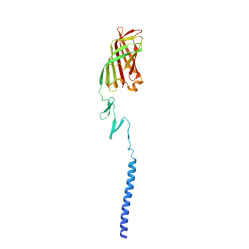
2VRS

Structure of avian reovirus sigmaC117-326, C2 crystal form


Experimental Data Snapshot

  • Method: X-RAY DIFFRACTION
  • Resolution: 1.75 Å
  • R-Value Free: 0.218 
  • R-Value Work: 0.180 
  • R-Value Observed: 0.180 

wwPDB Validation   3D Report Full Report


This is version 1.4 of the entry. See complete history


Literature

Crystallographic structure of the alpha-helical triple coiled-coil domain of avian reovirus S1133 fibre.

Guardado-Calvo, P.Fox, G.C.Llamas-Saiz, A.L.van Raaij, M.J.

(2009) J Gen Virol 90: 672-677

  • DOI: https://doi.org/10.1099/vir.0.008276-0
  • Primary Citation of Related Structures:  
    2JJL, 2VRS

  • PubMed Abstract: 

    Avian reovirus fibre, a homo-trimer of the sigmaC protein, is a minor component of the avian reovirus outer capsid. It is anchored via a short N-terminal sequence to the inner capsid lambdaC pentamer, and its protruding globular C-terminal domain is responsible for primary host cell attachment. We have previously solved the structure of a receptor-binding fragment in which residues 160-191 form a triple beta-spiral and 196-326 a beta-barrel head domain. Here we have expressed, purified and crystallized a major sigmaC fragment comprising residues 117-326. Its structure, which was solved by molecular replacement using the previously determined receptor-binding domain structure and refined to 1.75 A (0.175 nm) resolution, reveals an alpha-helical triple coiled-coil connected to the previously solved structure by a zinc-ion-containing linker. The coiled-coil domain contains two chloride ion binding sites, as well as specific trimerization and registration sequences. The linker may act as a functionally important hinge.


  • Organizational Affiliation

    Departamento de Bioquímica e Bioloxía Molecular, Facultade de Farmacia, Universidade de Santiago de Compostela, E-15782 Santiago de Compostela, Spain.


Macromolecules
Find similar proteins by:  (by identity cutoff)  |  3D Structure
Entity ID: 1
MoleculeChains Sequence LengthOrganismDetailsImage
SIGMA-C CAPSID PROTEIN
A, B, C
211Avian orthoreovirusMutation(s): 0 
UniProt
Find proteins for Q992I2 (Avian reovirus (strain S1133))
Explore Q992I2 
Go to UniProtKB:  Q992I2
Entity Groups  
Sequence Clusters30% Identity50% Identity70% Identity90% Identity95% Identity100% Identity
UniProt GroupQ992I2
Sequence Annotations
Expand
  • Reference Sequence
Small Molecules
Ligands 4 Unique
IDChains Name / Formula / InChI Key2D Diagram3D Interactions
SO4
Query on SO4

Download Ideal Coordinates CCD File 
M [auth B]SULFATE ION
O4 S
QAOWNCQODCNURD-UHFFFAOYSA-L
GOL
Query on GOL

Download Ideal Coordinates CCD File 
D [auth A],
J [auth B],
K [auth B]
GLYCEROL
C3 H8 O3
PEDCQBHIVMGVHV-UHFFFAOYSA-N
ZN
Query on ZN

Download Ideal Coordinates CCD File 
E [auth A]
F [auth A]
G [auth A]
H [auth A]
I [auth A]
E [auth A],
F [auth A],
G [auth A],
H [auth A],
I [auth A],
L [auth B],
P [auth C]
ZINC ION
Zn
PTFCDOFLOPIGGS-UHFFFAOYSA-N
CL
Query on CL

Download Ideal Coordinates CCD File 
N [auth C],
O [auth C]
CHLORIDE ION
Cl
VEXZGXHMUGYJMC-UHFFFAOYSA-M
Experimental Data & Validation

Experimental Data

  • Method: X-RAY DIFFRACTION
  • Resolution: 1.75 Å
  • R-Value Free: 0.218 
  • R-Value Work: 0.180 
  • R-Value Observed: 0.180 
  • Space Group: C 1 2 1
Unit Cell:
Length ( Å )Angle ( ˚ )
a = 142.789α = 90
b = 54.008β = 101.79
c = 121.336γ = 90
Software Package:
Software NamePurpose
REFMACrefinement
HKL-2000data reduction
SCALEPACKdata scaling
MOLREPphasing

Structure Validation

View Full Validation Report


Entry History 

Deposition Data

Revision History  (Full details and data files)

  • Version 1.0: 2009-01-13
    Type: Initial release
  • Version 1.1: 2011-05-08
    Changes: Version format compliance
  • Version 1.2: 2011-07-13
    Changes: Version format compliance
  • Version 1.3: 2018-02-07
    Changes: Database references
  • Version 1.4: 2023-12-13
    Changes: Data collection, Database references, Derived calculations, Other, Refinement description