2PES

Urate Oxidase in complex with tris-dipicolinate Lutetium


Experimental Data Snapshot

  • Method: X-RAY DIFFRACTION
  • Resolution: 1.60 Å
  • R-Value Free: 0.195 
  • R-Value Work: 0.179 
  • R-Value Observed: 0.180 

wwPDB Validation   3D Report Full Report


Ligand Structure Quality Assessment 


This is version 1.3 of the entry. See complete history


Literature

A dipicolinate lanthanide complex for solving protein structures using anomalous diffraction.

Pompidor, G.Maury, O.Vicat, J.Kahn, R.

(2010) Acta Crystallogr D Biol Crystallogr 66: 762-769

  • DOI: https://doi.org/10.1107/S0907444910010954
  • Primary Citation of Related Structures:  
    2PE7, 2PES, 3LGR

  • PubMed Abstract: 

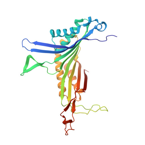
    Tris-dipicolinate lanthanide complexes were used to prepare derivative crystals of six proteins: hen egg-white lysozyme, turkey egg-white lysozyme, thaumatin from Thaumatococcus daniellii, urate oxidase from Aspergillus flavus, porcine pancreatic elastase and xylanase from Trichoderma reesei. Diffraction data were collected using either synchrotron radiation or X-rays from a laboratory source. In all cases, the complex turned out to be bound to the protein and the phases determined using the anomalous scattering of the lanthanide led to high-quality electron-density maps. The binding mode of the complex was characterized from the refined structures. The lanthanide tris-dipicolinate was found to bind through interactions between carboxylate groups of the dipicolinate ligands and hydrogen-bond donor groups of the protein. In each binding site, one enantiomeric form of the complex is selected from the racemic solution according to the specific site topology. For hen egg-white lysozyme and xylanase, derivative crystals obtained by cocrystallization belonged to a new monoclinic C2 crystal form that diffracted to high resolution.


  • Organizational Affiliation

    Institut de Biologie Structurale J.-P. Ebel, UMR 5075, 41 Rue Jules Horowitz, Grenoble, France.


Macromolecules
Find similar proteins by:  (by identity cutoff)  |  3D Structure
Entity ID: 1
MoleculeChains Sequence LengthOrganismDetailsImage
Uricase295Aspergillus flavusMutation(s): 0 
EC: 1.7.3.3
UniProt
Find proteins for Q00511 (Aspergillus flavus)
Explore Q00511 
Go to UniProtKB:  Q00511
Entity Groups  
Sequence Clusters30% Identity50% Identity70% Identity90% Identity95% Identity100% Identity
UniProt GroupQ00511
Sequence Annotations
Expand
  • Reference Sequence
Small Molecules
Ligands 3 Unique
IDChains Name / Formula / InChI Key2D Diagram3D Interactions
LU
Query on LU

Download Ideal Coordinates CCD File 
B [auth A],
C [auth A],
D [auth A]
LUTETIUM (III) ION
Lu
PSDMOPINLDTFSZ-UHFFFAOYSA-N
PDC
Query on PDC

Download Ideal Coordinates CCD File 
F [auth A]
G [auth A]
H [auth A]
I [auth A]
J [auth A]
F [auth A],
G [auth A],
H [auth A],
I [auth A],
J [auth A],
K [auth A]
PYRIDINE-2,6-DICARBOXYLIC ACID
C7 H5 N O4
WJJMNDUMQPNECX-UHFFFAOYSA-N
AZA
Query on AZA

Download Ideal Coordinates CCD File 
E [auth A]8-AZAXANTHINE
C4 H3 N5 O2
KVGVQTOQSNJTJI-UHFFFAOYSA-N
Experimental Data & Validation

Experimental Data

  • Method: X-RAY DIFFRACTION
  • Resolution: 1.60 Å
  • R-Value Free: 0.195 
  • R-Value Work: 0.179 
  • R-Value Observed: 0.180 
  • Space Group: I 2 2 2
Unit Cell:
Length ( Å )Angle ( ˚ )
a = 79.55α = 90
b = 95.32β = 90
c = 104.33γ = 90
Software Package:
Software NamePurpose
SCALAdata scaling
SHARPphasing
DMphasing
REFMACrefinement
PDB_EXTRACTdata extraction

Structure Validation

View Full Validation Report


Ligand Structure Quality Assessment 


Entry History 

Deposition Data

Revision History  (Full details and data files)

  • Version 1.0: 2008-04-22
    Type: Initial release
  • Version 1.1: 2011-07-13
    Changes: Derived calculations, Version format compliance
  • Version 1.2: 2012-03-21
    Changes: Database references
  • Version 1.3: 2024-02-21
    Changes: Data collection, Database references, Derived calculations