2P5K

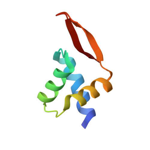
Crystal structure of the N-terminal domain of AhrC


Experimental Data Snapshot

  • Method: X-RAY DIFFRACTION
  • Resolution: 1.00 Å
  • R-Value Free: 0.160 
  • R-Value Work: 0.137 
  • R-Value Observed: 0.139 

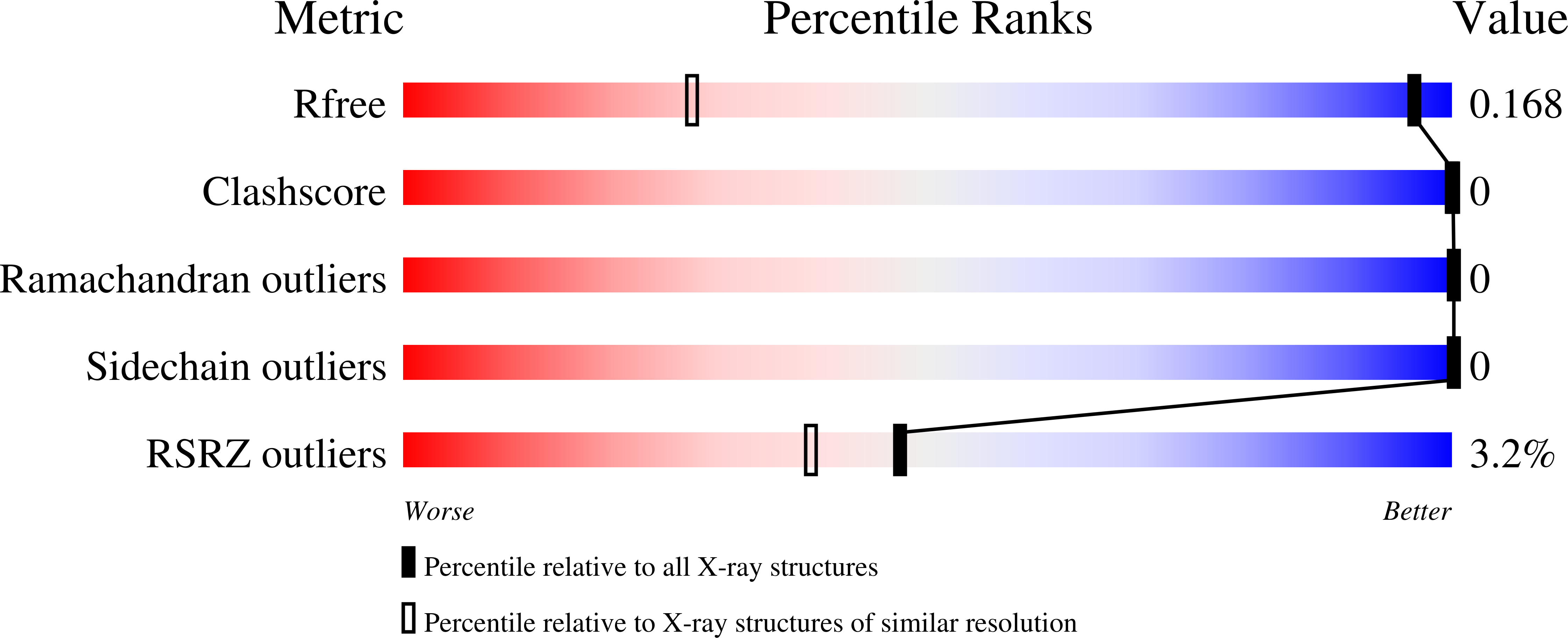
wwPDB Validation   3D Report Full Report


This is version 1.2 of the entry. See complete history


Literature

A high-resolution structure of the DNA-binding domain of AhrC, the arginine repressor/activator protein from Bacillus subtilis.

Garnett, J.A.Baumberg, S.Stockley, P.G.Phillips, S.E.

(2007) Acta Crystallogr Sect F Struct Biol Cryst Commun 63: 914-917

  • DOI: https://doi.org/10.1107/S1744309107048166
  • Primary Citation of Related Structures:  
    2P5K

  • PubMed Abstract: 

    In Bacillus subtilis the concentration of L-arginine is controlled by the transcriptional regulator AhrC, which interacts with 18 bp DNA operator sites called ARG boxes in the promoters of arginine biosynthetic and catabolic operons. AhrC is a 100 kDa homohexamer, with each subunit having two domains. The C-terminal domains form the core, mediating intersubunit interactions and binding of the co-repressor L-arginine, whilst the N-terminal domains contain a winged helix-turn-helix DNA-binding motif and are arranged around the periphery. The N-terminal domain of AhrC has been expressed, purified and characterized and it has been shown that the fragment still binds DNA operators as a recombinant monomer. The DNA-binding domain has also been crystallized and the crystal structure refined to 1.0 A resolution is presented.


  • Organizational Affiliation

    Astbury Centre for Structural Molecular Biology, University of Leeds, Leeds LS2 9JT, England.


Macromolecules
Find similar proteins by:  (by identity cutoff)  |  3D Structure
Entity ID: 1
MoleculeChains Sequence LengthOrganismDetailsImage
Arginine repressor64Bacillus subtilisMutation(s): 0 
Gene Names: argRahrC
UniProt
Find proteins for P17893 (Bacillus subtilis (strain 168))
Explore P17893 
Go to UniProtKB:  P17893
Entity Groups  
Sequence Clusters30% Identity50% Identity70% Identity90% Identity95% Identity100% Identity
UniProt GroupP17893
Sequence Annotations
Expand
  • Reference Sequence
Experimental Data & Validation

Experimental Data

  • Method: X-RAY DIFFRACTION
  • Resolution: 1.00 Å
  • R-Value Free: 0.160 
  • R-Value Work: 0.137 
  • R-Value Observed: 0.139 
  • Space Group: P 21 21 21
Unit Cell:
Length ( Å )Angle ( ˚ )
a = 25.108α = 90
b = 32.9β = 90
c = 66.226γ = 90
Software Package:
Software NamePurpose
REFMACrefinement
PDB_EXTRACTdata extraction
ADSCdata collection
MOSFLMdata reduction
SCALAdata scaling
AMoREphasing

Structure Validation

View Full Validation Report


Entry History 

Deposition Data

Revision History  (Full details and data files)

  • Version 1.0: 2007-10-30
    Type: Initial release
  • Version 1.1: 2011-07-13
    Changes: Version format compliance
  • Version 1.2: 2023-08-30
    Changes: Data collection, Database references, Refinement description