Multiple Conformations of the Cytidine Repressor DNA-Binding Domain Coalesce to One upon Recognition of a Specific DNA Surface.
Moody, C.L., Tretyachenko-Ladokhina, V., Laue, T.M., Senear, D.F., Cocco, M.J.(2011) Biochemistry 50: 6622-6632
- PubMed: 21688840
- DOI: https://doi.org/10.1021/bi200205v
- Primary Citation of Related Structures:
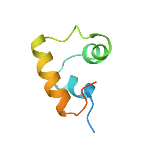
2L8N, 2LCV - PubMed Abstract:
The cytidine repressor (CytR) is a member of the LacR family of bacterial repressors with distinct functional features. The Escherichia coli CytR regulon comprises nine operons whose palindromic operators vary in both sequence and, most significantly, spacing between the recognition half-sites. This suggests a strong likelihood that protein folding would be coupled to DNA binding as a mechanism to accommodate the variety of different operator architectures to which CytR is targeted. Such coupling is a common feature of sequence-specific DNA-binding proteins, including the LacR family repressors; however, there are no significant structural rearrangements upon DNA binding within the three-helix DNA-binding domains (DBDs) studied to date. We used nuclear magnetic resonance (NMR) spectroscopy to characterize the CytR DBD free in solution and to determine the high-resolution structure of a CytR DBD monomer bound specifically to one DNA half-site of the uridine phosphorylase (udp) operator. We find that the free DBD populates multiple distinct conformations distinguished by up to four sets of NMR peaks per residue. This structural heterogeneity is previously unknown in the LacR family. These stable structures coalesce into a single, more stable udp-bound form that features a three-helix bundle containing a canonical helix-turn-helix motif. However, this structure differs from all other LacR family members whose structures are known with regard to the packing of the helices and consequently their relative orientations. Aspects of CytR activity are unique among repressors; we identify here structural properties that are also distinct and that might underlie the different functional properties.
Organizational Affiliation:
Department of Molecular Biology and Biochemistry, University of California, Irvine, California 92697, USA.