2HTT

Ruthenium Hexammine ion interactions with Z-DNA


Experimental Data Snapshot

  • Method: X-RAY DIFFRACTION
  • Resolution: 2.60 Å
  • R-Value Free: 0.364 
  • R-Value Work: 0.258 
  • R-Value Observed: 0.267 

wwPDB Validation   3D Report Full Report


This is version 1.4 of the entry. See complete history


Literature

Hexammineruthenium(III) ion interactions with Z-DNA

Bharanidharan, D.Thiyagarajan, S.Gautham, N.

(2007) Acta Crystallogr Sect F Struct Biol Cryst Commun 63: 1008-1013

  • DOI: https://doi.org/10.1107/S1744309107047781
  • Primary Citation of Related Structures:  
    2HTO, 2HTT

  • PubMed Abstract: 

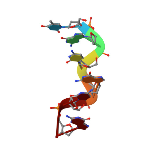
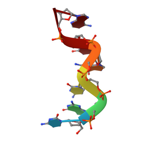
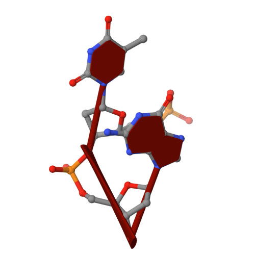
    The hexamer duplex d(CGCGCA).d(TGCGCG) was crystallized with hexammineruthenium(III) ions in an orthorhombic space group; the crystals diffracted to 1.54 A resolution. Strong ion interactions with the adenine base induce a tautomeric shift from the amino to the imino form. Consequently, the A.T base pairing is disrupted. This structural study may be relevant to metal toxicity.


  • Organizational Affiliation

    Department of Crystallography and Biophysics, University of Madras, Guindy Campus, Chennai 600 025, Tamil Nadu, India.


Macromolecules

Find similar nucleic acids by:  Sequence   |   3D Structure  

Entity ID: 1
MoleculeChains LengthOrganismImage
DNA (5'-D(*DTP*DGP*DCP*DGP*DCP*DG)-3')6N/A
Sequence Annotations
Expand
  • Reference Sequence

Find similar nucleic acids by:  Sequence   |   3D Structure  

Entity ID: 2
MoleculeChains LengthOrganismImage
DNA (5'-D(*DCP*DGP*DCP*DGP*DCP*DA)-3')6N/A
Sequence Annotations
Expand
  • Reference Sequence

Find similar nucleic acids by:  Sequence   |   3D Structure  

Entity ID: 3
MoleculeChains LengthOrganismImage
DNA (5'-D(P*DTP*DG)-3')C [auth D],
D [auth E]
2N/A
Sequence Annotations
Expand
  • Reference Sequence
Small Molecules
Ligands 1 Unique
IDChains Name / Formula / InChI Key2D Diagram3D Interactions
NRU
Query on NRU

Download Ideal Coordinates CCD File 
E
RUTHENIUM (III) HEXAAMINE ION
H18 N6 Ru
HOOVQFBYNWLMGV-UHFFFAOYSA-N
Experimental Data & Validation

Experimental Data

  • Method: X-RAY DIFFRACTION
  • Resolution: 2.60 Å
  • R-Value Free: 0.364 
  • R-Value Work: 0.258 
  • R-Value Observed: 0.267 
  • Space Group: P 65
Unit Cell:
Length ( Å )Angle ( ˚ )
a = 35.895α = 90
b = 35.895β = 90
c = 44.604γ = 120
Software Package:
Software NamePurpose
REFMACrefinement
AUTOMARdata reduction
AMoREphasing

Structure Validation

View Full Validation Report


Entry History 

Deposition Data

Revision History  (Full details and data files)

  • Version 1.0: 2006-08-08
    Type: Initial release
  • Version 1.1: 2008-05-01
    Changes: Version format compliance
  • Version 1.2: 2011-07-13
    Changes: Version format compliance
  • Version 1.3: 2024-03-13
    Changes: Data collection, Database references, Derived calculations
  • Version 1.4: 2024-04-03
    Changes: Refinement description