2H8A

Structure of Microsomal Glutathione Transferase 1 in Complex with Glutathione


Experimental Data Snapshot

  • Method: ELECTRON CRYSTALLOGRAPHY
  • Resolution: 3.20 Å
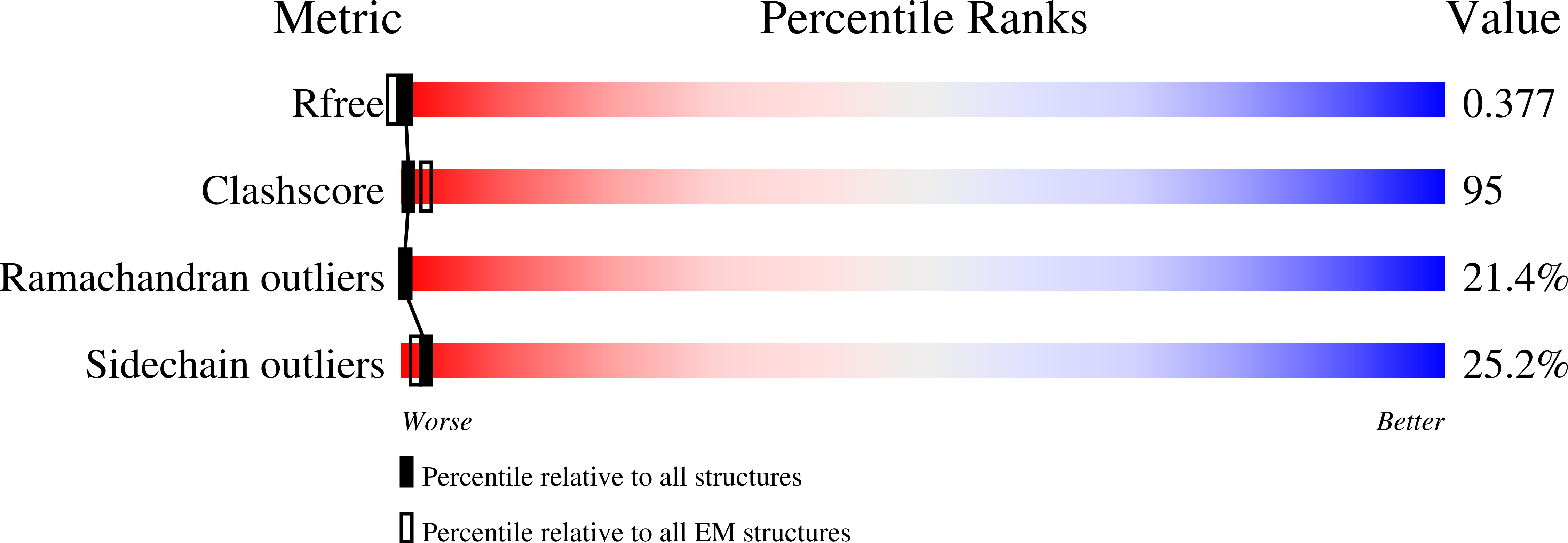
  • R-Value Free: 0.376 
  • R-Value Work: 0.339 
  • R-Value Observed: 0.342 

wwPDB Validation   3D Report Full Report


This is version 1.5 of the entry. See complete history


Literature

Structural basis for detoxification and oxidative stress protection in membranes.

Holm, P.J.Bhakat, P.Jegerschold, C.Gyobu, N.Mitsuoka, K.Fujiyoshi, Y.Morgenstern, R.Hebert, H.

(2006) J Mol Biol 360: 934-945

  • DOI: https://doi.org/10.1016/j.jmb.2006.05.056
  • Primary Citation of Related Structures:  
    2H8A

  • PubMed Abstract: 

    Synthesis of mediators of fever, pain and inflammation as well as protection against reactive molecules and oxidative stress is a hallmark of the MAPEG superfamily (membrane associated proteins in eicosanoid and glutathione metabolism). The structure of a MAPEG member, rat microsomal glutathione transferase 1, at 3.2 A resolution, solved here in complex with glutathione by electron crystallography, defines the active site location and a cytosolic domain involved in enzyme activation. The glutathione binding site is found to be different from that of the canonical soluble glutathione transferases. The architecture of the homotrimer supports a catalytic mechanism involving subunit interactions and reveals both cytosolic and membraneous substrate entry sites, providing a rationale for the membrane location of the enzyme.


  • Organizational Affiliation

    Department of Biosciences and Nutrition, Karolinska Institutet and School of Technology and Health, Royal Institute of Technology, SE-14157 Huddinge, Sweden.


Macromolecules
Find similar proteins by:  (by identity cutoff)  |  3D Structure
Entity ID: 1
MoleculeChains Sequence LengthOrganismDetailsImage
Microsomal glutathione S-transferase 1154Rattus norvegicusMutation(s): 0 
EC: 2.5.1.18
Membrane Entity: Yes 
UniProt
Find proteins for P08011 (Rattus norvegicus)
Explore P08011 
Go to UniProtKB:  P08011
Entity Groups  
Sequence Clusters30% Identity50% Identity70% Identity90% Identity95% Identity100% Identity
UniProt GroupP08011
Sequence Annotations
Expand
  • Reference Sequence
Small Molecules
Ligands 1 Unique
IDChains Name / Formula / InChI Key2D Diagram3D Interactions
GSH
Query on GSH

Download Ideal Coordinates CCD File 
B [auth A]GLUTATHIONE
C10 H17 N3 O6 S
RWSXRVCMGQZWBV-WDSKDSINSA-N
Experimental Data & Validation

Experimental Data

  • Method: ELECTRON CRYSTALLOGRAPHY
  • Resolution: 3.20 Å
  • R-Value Free: 0.376 
  • R-Value Work: 0.339 
  • R-Value Observed: 0.342 
  • Space Group: P 6
Unit Cell:
Length ( Å )Angle ( ˚ )
a = 81.8α = 90
b = 81.8β = 90
c = 100γ = 120
Software Package:
Software NamePurpose
REFMACrefinement

Structure Validation

View Full Validation Report


Entry History 

Deposition Data

  • Released Date: 2007-05-22 
  • Deposition Author(s): Hebert, H.

Revision History  (Full details and data files)

  • Version 1.0: 2007-05-22
    Type: Initial release
  • Version 1.1: 2008-05-01
    Changes: Version format compliance
  • Version 1.2: 2011-07-13
    Changes: Derived calculations, Version format compliance
  • Version 1.3: 2012-01-18
    Changes: Non-polymer description
  • Version 1.4: 2012-03-21
    Changes: Non-polymer description
  • Version 1.5: 2024-02-14
    Changes: Author supporting evidence, Data collection, Database references, Derived calculations