Crystal structures of SnoaL2 and AclR: two putative hydroxylases in the biosynthesis of aromatic polyketide antibiotics
Beinker, P., Lohkamp, B., Peltonen, T., Niemi, J., Mantsala, P., Schneider, G.(2006) J Mol Biol 359: 728-740
- PubMed: 16650858
- DOI: https://doi.org/10.1016/j.jmb.2006.03.060
- Primary Citation of Related Structures:
2GEX, 2GEY - PubMed Abstract:
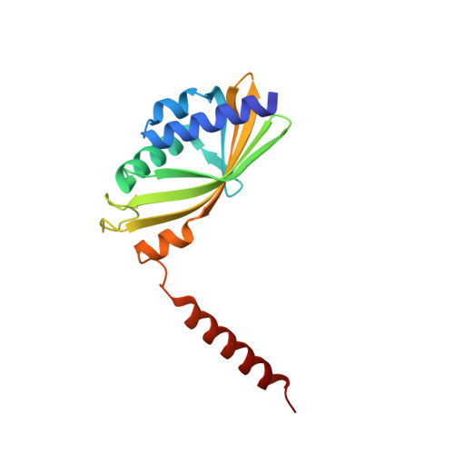
SnoaL2 and AclR are homologous enzymes in the biosynthesis of the aromatic polyketides nogalamycin in Streptomyces nogalater and cinerubin in Streptomyces galilaeus, respectively. Evidence obtained from gene transfer experiments suggested that SnoaL2 catalyzes the hydroxylation of the C-1 carbon atom of the polyketide chain. Here we show that AclR is also involved in the production of 1-hydroxylated anthracyclines in vivo. The three-dimensional structure of SnoaL2 has been determined by multi-wavelength anomalous diffraction to 2.5A resolution, and that of AclR to 1.8A resolution using molecular replacement. Both enzymes are dimers in solution and in the crystal. The fold of the enzyme subunits consists of an alpha+beta barrel. The dimer interface is formed by packing of the beta-sheets from the two subunits against each other. In the interior of the alpha+beta barrel a hydrophobic cavity is formed that most likely binds the substrate and harbors the active site. The subunit fold and the architecture of the active site in SnoaL2 and AclR are similar to that of the polyketide cyclases SnoaL and AknH; however, they show completely different quaternary structures. A comparison of the active site pockets of the putative hydroxylases AclR and SnoaL2 with those of bona fide polyketide cyclases reveals distinct differences in amino acids lining the cavity that might be responsible for the switch in chemistry. The moderate degree of sequence similarity and the preservation of the three-dimensional fold of the polypeptide chain suggest that these enzymes are evolutionary related. Members of this enzyme family appear to have evolved from a common protein scaffold by divergent evolution to catalyze reactions chemically as diverse as aldol condensation and hydroxylation.
Organizational Affiliation:
Department of Medical Biochemistry and Biophysics, Karolinska Institutet, Stockholm S-171 77, Sweden.