2GB0

Monomeric sarcosine oxidase: structure of a covalently flavinylated amine oxidizing enzyme


Experimental Data Snapshot

  • Method: X-RAY DIFFRACTION
  • Resolution: 1.85 Å
  • R-Value Free: 0.198 
  • R-Value Work: 0.165 

wwPDB Validation   3D Report Full Report


Ligand Structure Quality Assessment 


This is version 1.3 of the entry. See complete history


Literature

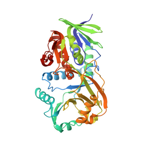
Monomeric Sarcosine Oxidase: Structure of a Covalently Flavinylated Amine Oxidizing Enzyme

Trickey, P.Wagner, M.A.Jorns, M.S.Mathews, F.S.

(1999) Structure 7: 331-345

  • DOI: https://doi.org/10.1016/s0969-2126(99)80043-4
  • Primary Citation of Related Structures:  
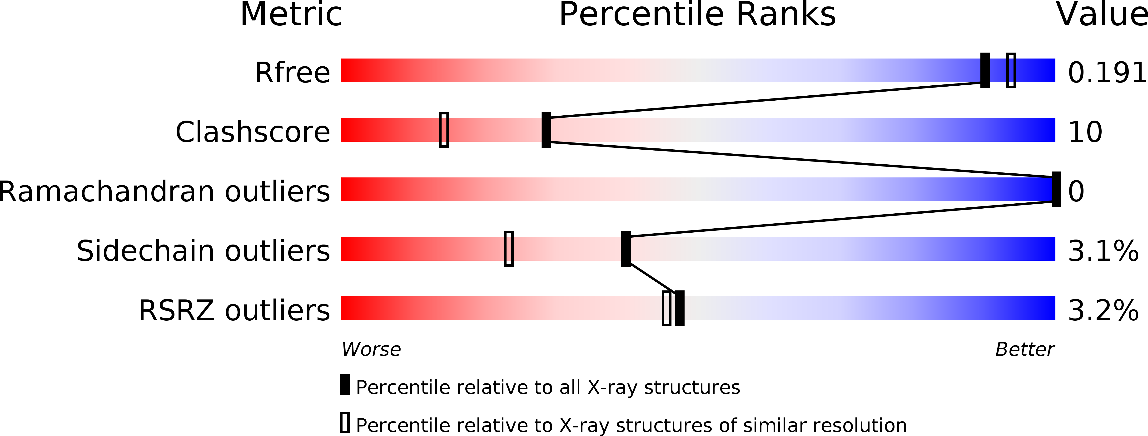
    2GB0

  • PubMed Abstract: 

    Monomeric sarcosine oxidases (MSOXs) are among the simplest members of a recently recognized family of eukaryotic and prokaryotic enzymes that catalyze similar oxidative reactions with various secondary or tertiary amino acids and contain covalently bound flavins. Other members of this family include heterotetrameric sarcosine oxidase, N-methyltryptophan oxidase and pipecolate oxidase. Mammalian sarcosine dehydrogenase and dimethylglycine dehydrogenase may be more distantly related family members. The X-ray crystal structure of MSOX from Bacillus sp. B-0618, expressed in Escherichia coli, has been solved at 2.0 A resolution by multiwavelength anomalous dispersion (MAD) from crystals of the selenomethionine-substituted enzyme. Fourteen selenium sites, belonging to two MSOX molecules in the asymmetric unit, were used for MAD phasing and to define the local twofold symmetry axis for electron-density averaging. The structures of the native enzyme and of two enzyme-inhibitor complexes were also determined. MSOX is a two-domain protein with an overall topology most similar to that of D-amino acid oxidase, with which it shares 14% sequence identity. The flavin ring is located in a very basic environment, making contact with sidechains of arginine, lysine, histidine and the N-terminal end of a helix dipole. The flavin is covalently attached through an 8alpha-S-cysteinyl linkage to Cys315 of the catalytic domain. Covalent attachment is probably self-catalyzed through interactions with the positive sidechains and the helix dipole. Substrate binding is probably stabilized by hydrogen bonds between the substrate carboxylate and two basic sidechains, Arg52 and Lys348, located above the re face of the flavin ring.


  • Organizational Affiliation

    Department of Biochemistry and Molecular Biophysics, Washington University School of Medicine, 660 S. Euclid Ave, St. Louis, MO 63110, USA.


Macromolecules
Find similar proteins by:  (by identity cutoff)  |  3D Structure
Entity ID: 1
MoleculeChains Sequence LengthOrganismDetailsImage
Monomeric sarcosine oxidase
A, B
389Bacillus sp. B-0618Mutation(s): 0 
Gene Names: soxAsox
EC: 1.5.3.1
UniProt
Find proteins for P40859 (Bacillus sp. (strain B-0618))
Explore P40859 
Go to UniProtKB:  P40859
Entity Groups  
Sequence Clusters30% Identity50% Identity70% Identity90% Identity95% Identity100% Identity
UniProt GroupP40859
Sequence Annotations
Expand
  • Reference Sequence
Small Molecules
Ligands 3 Unique
IDChains Name / Formula / InChI Key2D Diagram3D Interactions
FAD
Query on FAD

Download Ideal Coordinates CCD File 
E [auth A],
J [auth B]
FLAVIN-ADENINE DINUCLEOTIDE
C27 H33 N9 O15 P2
VWWQXMAJTJZDQX-UYBVJOGSSA-N
PO4
Query on PO4

Download Ideal Coordinates CCD File 
C [auth A],
F [auth B],
G [auth B],
H [auth B]
PHOSPHATE ION
O4 P
NBIIXXVUZAFLBC-UHFFFAOYSA-K
CL
Query on CL

Download Ideal Coordinates CCD File 
D [auth A],
I [auth B]
CHLORIDE ION
Cl
VEXZGXHMUGYJMC-UHFFFAOYSA-M
Experimental Data & Validation

Experimental Data

  • Method: X-RAY DIFFRACTION
  • Resolution: 1.85 Å
  • R-Value Free: 0.198 
  • R-Value Work: 0.165 
  • Space Group: P 1 21 1
Unit Cell:
Length ( Å )Angle ( ˚ )
a = 72.562α = 90
b = 69.555β = 94.26
c = 74.117γ = 90
Software Package:
Software NamePurpose
DENZOdata reduction
SCALEPACKdata scaling
SOLVEphasing
SHARPphasing
DMmodel building
CNSrefinement
DMphasing

Structure Validation

View Full Validation Report


Ligand Structure Quality Assessment 


Entry History 

Deposition Data

Revision History  (Full details and data files)

  • Version 1.0: 2006-03-21
    Type: Initial release
  • Version 1.1: 2008-05-01
    Changes: Version format compliance
  • Version 1.2: 2011-07-13
    Changes: Source and taxonomy, Version format compliance
  • Version 1.3: 2017-10-18
    Changes: Refinement description