2DFC

Xylanase II from Tricoderma reesei at 293K


Experimental Data Snapshot

  • Method: X-RAY DIFFRACTION
  • Resolution: 1.19 Å
  • R-Value Free: 0.122 
  • R-Value Work: 0.106 
  • R-Value Observed: 0.106 

wwPDB Validation   3D Report Full Report


This is version 2.1 of the entry. See complete history

Re-refinement Note

A newer entry is available that reflects an alternative modeling of the original data: 6JUG 6JWB 6JXL 6K9O 6K9R


Literature

Structure of an orthorhombic form of xylanase II from Trichoderma reesei and analysis of thermal displacement.

Watanabe, N.Akiba, T.Kanai, R.Harata, K.

(2006) Acta Crystallogr D Biol Crystallogr 62: 784-792

  • DOI: https://doi.org/10.1107/S0907444906017379
  • Primary Citation of Related Structures:  
    2DFB, 2DFC

  • PubMed Abstract: 

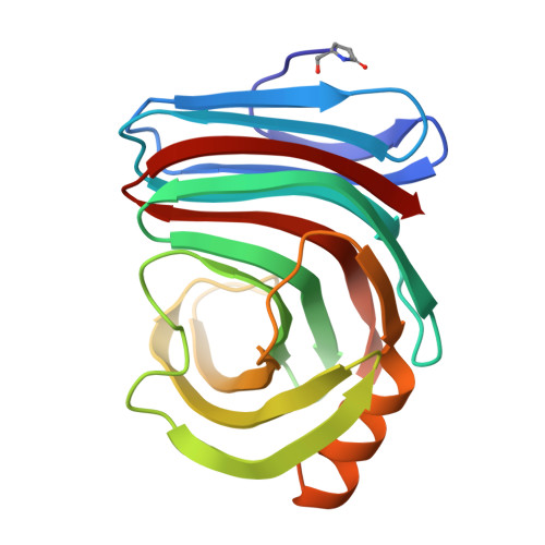
    An orthorhombic crystal of xylanase II from Trichoderma reesei was grown in the presence of sodium iodide. Crystal structures at atomic resolution were determined at 100 and 293 K. Protein molecules were aligned along a crystallographic twofold screw axis, forming a helically extended polymer-like chain mediated by an iodide ion. The iodide ion connected main-chain peptide groups between two adjacent molecules by an N-H...I-...H-N hydrogen-bond bridge, thus contributing to regulation of the molecular arrangement and suppression of the rigid-body motion in the crystal with high diffraction quality. The structure at 293 K showed considerable thermal motion in the loop regions connecting the beta-strands that form the active-site cleft. TLS model analysis of the thermal motion and a comparison between this structure and that at 100 K suggest that the fluctuation of these loop regions is attributable to the hinge-like movement of the beta-strands.


  • Organizational Affiliation

    Biological Information Research Center, National Institute of Advanced Industrial Science and Technology (AIST), Central 6, 1-1-1 Higashi, Tsukuba, Ibaraki 305-8566, Japan.


Macromolecules
Find similar proteins by:  (by identity cutoff)  |  3D Structure
Entity ID: 1
MoleculeChains Sequence LengthOrganismDetailsImage
Endo-1,4-beta-xylanase 2190Trichoderma reeseiMutation(s): 0 
EC: 3.2.1.8
UniProt
Find proteins for P36217 (Hypocrea jecorina (strain ATCC 56765 / BCRC 32924 / NRRL 11460 / Rut C-30))
Explore P36217 
Go to UniProtKB:  P36217
Entity Groups  
Sequence Clusters30% Identity50% Identity70% Identity90% Identity95% Identity100% Identity
UniProt GroupP36217
Sequence Annotations
Expand
  • Reference Sequence
Small Molecules
Ligands 1 Unique
IDChains Name / Formula / InChI Key2D Diagram3D Interactions
IOD
Query on IOD

Download Ideal Coordinates CCD File 
B [auth A]IODIDE ION
I
XMBWDFGMSWQBCA-UHFFFAOYSA-M
Modified Residues  1 Unique
IDChains TypeFormula2D DiagramParent
PCA
Query on PCA
A
L-PEPTIDE LINKINGC5 H7 N O3GLN
Experimental Data & Validation

Experimental Data

  • Method: X-RAY DIFFRACTION
  • Resolution: 1.19 Å
  • R-Value Free: 0.122 
  • R-Value Work: 0.106 
  • R-Value Observed: 0.106 
  • Space Group: P 21 21 21
Unit Cell:
Length ( Å )Angle ( ˚ )
a = 49.55α = 90
b = 60.03β = 90
c = 70.5γ = 90
Software Package:
Software NamePurpose
HKL-2000data collection
TRUNCATEdata reduction
CNSrefinement
SHELXL-97refinement
HKL-2000data reduction
CCP4data scaling
CNSphasing

Structure Validation

View Full Validation Report


Entry History 

Deposition Data

Revision History  (Full details and data files)

  • Version 1.0: 2006-07-11
    Type: Initial release
  • Version 1.1: 2008-04-30
    Changes: Version format compliance
  • Version 1.2: 2011-07-13
    Changes: Version format compliance
  • Version 2.0: 2019-12-25
    Changes: Database references, Derived calculations, Polymer sequence
  • Version 2.1: 2023-10-25
    Changes: Data collection, Database references, Derived calculations, Refinement description